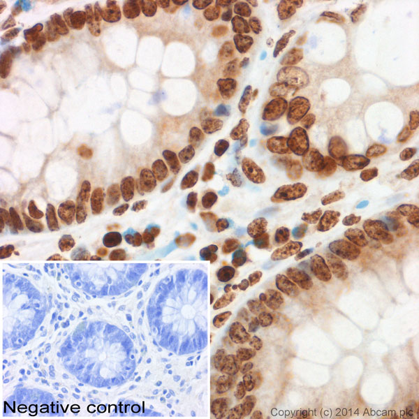
IHC image of ab8898 staining Histone H3 (tri methyl K9) in normal human colon formalin-fixed paraffin-embedded tissue sections*, performed on a Leica Bond. The section was pre-treated using heat mediated antigen retrieval with sodium citrate buffer (pH6, epitope retrieval solution 1) for 20 mins. The section was then incubated with ab8898, 1/400 dilution, for 15 mins at room temperature and detected using an HRP conjugated compact polymer system. DAB was used as the chromogen. The section was then counterstained with haematoxylin and mounted with DPX. No primary antibody was used in the negative control (shown on the inset).For other IHC staining systems (automated and non-automated) customers should optimize variable parameters such as antigen retrieval conditions, primary antibody concentration and antibody incubation times.*Tissue obtained from the Human Research Tissue Bank, supported by the NIHR Cambridge Biomedical Research Centre
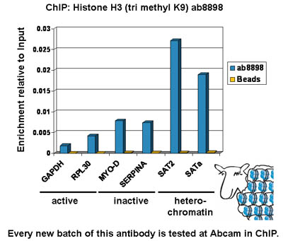
Chromatin was prepared from U2OS cells according to the Abcam X-ChIP protocol. Cells were fixed with formaldehyde for 10 min. The ChIP was performed with 25 µg of chromatin, 2 µg of ab8898 (blue), and 20 µl of protein A/G sepharose beads. No antibody was added to the beads control (yellow). The immunoprecipitated DNA was quantified by real time PCR (Taqman approach for active and inactive loci, Sybr green approach for heterochromatic loci). Primers and probes are located in the first kb of the transcribed region.
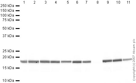
All lanes : Anti-Histone H3 (tri methyl K9) antibody - ChIP Grade (ab8898) at 1 µg/mlLane 1 : Calf Thymus Histone Preparation Nuclear LysateLane 2 : Calf Thymus Histone Preparation Nuclear Lysate with Human Histone H3 (unmodified ) peptide (ab7228) at 0.5 µg/mlLane 3 : Calf Thymus Histone Preparation Nuclear Lysate with Human Histone H3 (mono methyl K4) peptide (ab1340) at 0.5 µg/mlLane 4 : Calf Thymus Histone Preparation Nuclear Lysate with Human Histone H3 (di methyl K4) peptide (ab7768) at 0.5 µg/mlLane 5 : Calf Thymus Histone Preparation Nuclear Lysate with Human Histone H3 (tri methyl K4) peptide (ab1342) at 0.5 µg/mlLane 6 : Calf Thymus Histone Preparation Nuclear Lysate with Human Histone H3 (mono methyl K9) peptide (ab1771) at 0.5 µg/mlLane 7 : Calf Thymus Histone Preparation Nuclear Lysate with Human Histone H3 (di methyl K9) peptide (ab1772) at 0.5 µg/mlLane 8 : Calf Thymus Histone Preparation Nuclear Lysate with Human Histone H3 (tri methyl K9) peptide (ab1773) at 0.5 µg/mlL
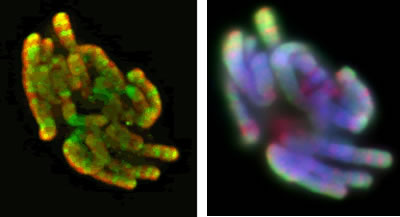
Indian muntjac fibroblast cells stained with anti-Histone H3 tri methyl K9, ab8898, (green, left panel, deconvolution image; red, right panel, epifluorescence image).The centromeres are enriched in Histone H3 tri methyl K9. There are also additional bands that occur throughout the chromosomes. Note that these images are taken in situ and are imaged under conditions where distinct cytogenetic-like banding patterns have not previously been possible to visualize (e.g., several acetylated antibodies have been reported to be associated with chromosome bands but, although not homogenously distributed along in situ chromosomes, they do not generate distinct banding patterns).
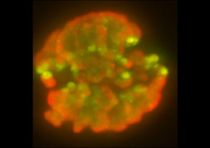
A 3-D reconstruction of a mouse embryonic fibroblast cell in metaphase stained with anti-Histone H3 tri methyl K9(green, ab8898) and DAPI (red).

ab8898 staining human uterine tumour tissue sections by IHC-P. Sections were fixed in formaldehyde and subjected to heat mediated antigen retrieval in citrate buffer (pH 6) prior to blocking with 5% serum for 30 minutes at 22°C. The primary antibody was diluted 1/400 and incubated with the sample for 30 minutes at 22°C. A HRP-conjugated goat anti-rabbit antibody diluted 1/400, was used as the secondary.See Abreview
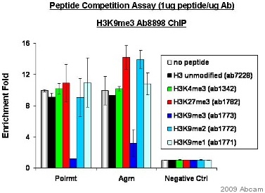
X-Chip assay was performed using nuclear lysates prepared from mouse ES cells. Crosslinking was done for 15 minutes in 1% formaldehyde. Primary antibody was incubated first with peptides ab7228, ab1342, ab1782, ab1773, ab1772 and ab1771 in a chip competition assay and then used in chip at 0.0133µg/ µg chromatin (chip sonication buffer) and incubated with sample for 24 hours at 4°C.Positive control: ChIP coupled with a peptide competition assay to validate the specificity of the antibody.Negative control: Genomic region (chr10:79154149-79155200) with no evidence of H3K9me3. RT-PCR detection method was used.Polrmt: PCR primers situated in the coding regions of Polymerase (RNA) mitochondrial (DNA directed).Agrn: PCR primers situated in the coding regions of Agrin
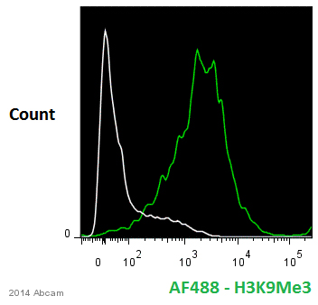
ab8898 staining Histone H3 (tri methyl K9) in human differentiated haematopoietic stem cells by Flow Cytometry. Cells were fixed with paraformaldehyde and permeabilized with permeabilization buffer. The sample was incubated with the primary antibody (1/600) for 12 hours at 4°C. An Alexa Fluor® 488-conjugated goat polyclonal anti-rabbit IgG (1/1000) was used as the secondary antibody.Gating Strategy: Isotype negative control (white).See Abreview
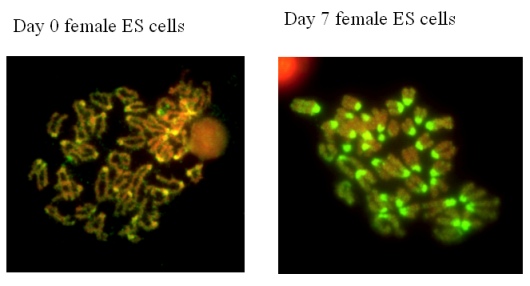
These images were kindly submitted by Prof Bryan Turner, University of Birmingham. Undifferentiated Mouse Embryonic Stem cells or cells differentiated for 7 days were incubated with ab8898. The staining is specific for centromeric heterochromatin on metaphase chromosomes.
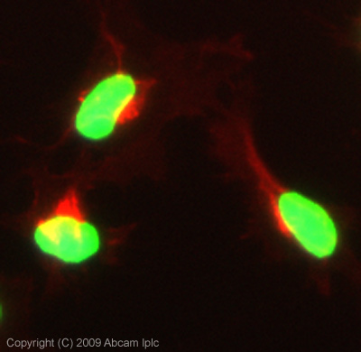
ICC/IF image of ab8898 stained HeLa cells. The cells were 100% methanol fixed (5 min) and then incubated in 1%BSA / 10% normal goat serum / 0.3M glycine in 0.1% PBS-Tween for 1h to permeabilise the cells and block non-specific protein-protein interactions. The cells were then incubated with the antibody (ab8898, 0.1µg/ml) overnight at +4°C. The secondary antibody (green) was Alexa Fluor® 488 goat anti-rabbit IgG (H+L) used at a 1/1000 dilution for 1h. Alexa Fluor® 594 WGA was used to label plasma membranes (red) at a 1/200 dilution for 1h. DAPI was used to stain the cell nuclei (blue) at a concentration of 1.43µM. This antibody also gave a positive result in 100% methanol fixed (5 min) HepG2 and MCF7 cells at 0.1µg/ml and in 4% PFA fixed (10 min) HeLa, Hek293, HepG2 and MCF7 cells at 0.1ug/ml.
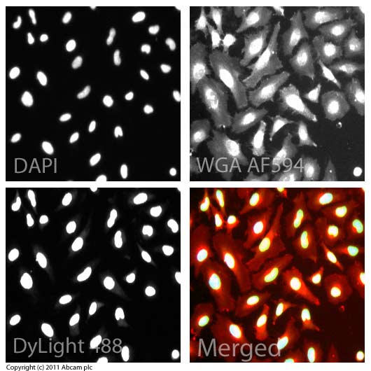
ICC/IF image of ab8898 stained HeLa cells. The cells were 100% methanol fixed (5 min) and then incubated in 1%BSA / 10% normal goat serum / 0.3M glycine in 0.1% PBS-Tween for 1h to permeabilise the cells and block non-specific protein-protein interactions. The cells were then incubated with the antibody (ab8898, 0.1µg/ml) overnight at +4°C. The secondary antibody (green) was ab96899, a goat anti-rabbit DyLight® 488 (IgG; H+L) used at a 1/250 dilution for 1h. Alexa Fluor® 594 WGA was used to label plasma membranes (red) at a 1/200 dilution for 1h. DAPI was used to stain the cell nuclei (blue) at a concentration of 1.43µM.
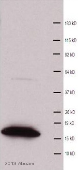
Anti-Histone H3 (tri methyl K9) antibody - ChIP Grade (ab8898) at 1/500 dilution + Rat E13 embryonic brain tissue lysate - nuclear at 10 µgSecondaryHRP-conjugated Goat anti-rabbit IgG polyclonal at 1/2000 dilutiondeveloped using the ECL techniquePerformed under reducing conditions.
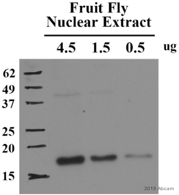
All lanes : Anti-Histone H3 (tri methyl K9) antibody - ChIP Grade (ab8898) at 1/1000 dilutionLane 1 : Fruit fly embryo tissue lysate - nuclear at 4.5 µgLane 2 : Fruit fly embryo tissue lysate - nuclear at 1.5 µgLane 3 : Fruit fly embryo tissue lysate - nuclear at 0.5 µgSecondaryHRP-conjugated Goat anti-rabbit IgG polyclonal at 1/2000 dilutiondeveloped using the ECL techniquePerformed under reducing conditions.


