Anti-Histone H3 (mono methyl R2) antibody - ChIP Grade
| Name | Anti-Histone H3 (mono methyl R2) antibody - ChIP Grade |
|---|---|
| Supplier | Abcam |
| Catalog | ab15584 |
| Prices | $401.00 |
| Sizes | 50 µg |
| Host | Rabbit |
| Clonality | Polyclonal |
| Isotype | IgG |
| Applications | IP ChIP ChIP DB IHC-P Array WB ChIP |
| Species Reactivities | Mouse, Bovine, Human, Yeast |
| Antigen | Synthetic peptide conjugated to KLH derived from within residues 1 to the C-terminus Histone H3, mono methylated at R2 |
| Description | Rabbit Polyclonal |
| Gene | HIST3H3 |
| Conjugate | Unconjugated |
| Supplier Page | Shop |
Product images
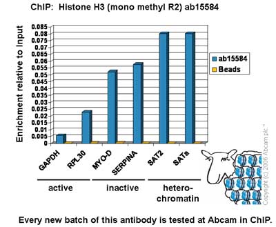 Chromatin was prepared from Hela cells according to the Abcam X-ChIP protocol. Cells were fixed with formaldehyde for 10min. The ChIP was performed with 25µg of chromatin, 2µg of ab15584 (blue), and 20µl of Protein A/G sepharose beads. No antibody was added to the beads control (yellow). The immunoprecipitated DNA was quantified by real time PCR (Taqman approach). Primers and probes are located in the first kb of the transcribed region.
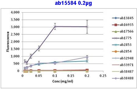 All batches of ab15584 are tested in Peptide Array against peptides to different Histone H3 modifications. Six dilutions of each peptide are printed on to the Peptide Array in triplicate and results are averaged before being plotted on to a graph. Results show strong binding to Histone H3 - mono methyl R2 peptide (ab1775), indicating that this antibody specifically recognises the Histone H3 - mono methyl R2 modification.1. ab1775 – Histone H3 - mono methyl R2 peptide2. ab38488 – Histone H3 - symmetric di methyl R23. ab2853 – Histone H3 - asymmetric di methyl R24. ab17566 – Histone H3 - non-modified5. ab33971 – Histone H3 - mono methyl R86. ab13845 – Histone H3 - mono methyl R177. ab16935 – Histone H3 - asymmetric di methyl R178. ab32948 – Histone H3 - symm
 Anti-Histone H3 (mono methyl R2) antibody - ChIP Grade (ab15584) at 1 µg/ml + Calf Thymus Histone Preparation Nuclear Lysate at 0.5 µgSecondaryGoat polyclonal to Rabbit IgG - H&L - Pre-Adsorbed (HRP) at 1/3000 dilutionPerformed under reducing conditions.
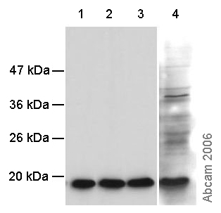 All lanes : Anti-Histone H3 (mono methyl R2) antibody - ChIP Grade (ab15584) at 0.5 µg/mlLane 1 : Calf thymus histone at 0.4 µgLane 2 : C3H10T1/2 histones at 0.4 µgLane 3 : 293 histones at 0.4 µgLane 4 : Yeast extracts from 1 x 10⁶ cells
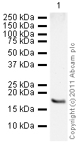
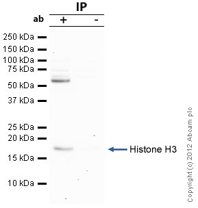 Histone H3 was immunoprecipitated using 0.5mg Hela whole cell extract, 5µg of Rabbit polyclonal to Histone H3 and 50µl of protein G magnetic beads (+). No antibody was added to the control (-). The antibody was incubated under agitation with Protein G beads for 10min, Hela whole cell extract lysate diluted in RIPA buffer was added to each sample and incubated for a further 10min under agitation.Proteins were eluted by addition of 40µl SDS loading buffer and incubated for 10min at 70oC; 10µl of each sample was separated on a SDS PAGE gel, transferred to a nitrocellulose membrane, blocked with 5% BSA and probed with ab15584.Secondary: Clean-Blot IP Detection Reagent (HRP) at 1/500 dilution.Band: 17kDa; Histone H3
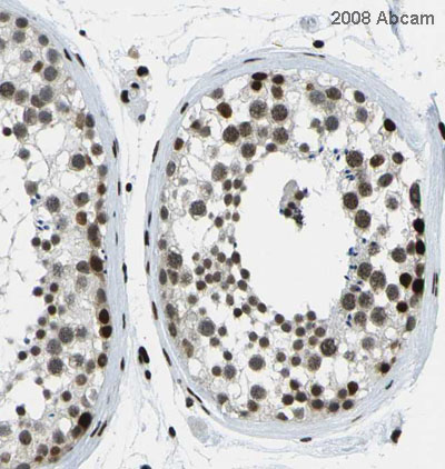 Image courtesy of Human Protein Atlas ab15584 staining histone H3 mono methyl R2 in human testis, showing a distinct and strong nuclear staining pattern in ductus seminiferus and leydig cells. Paraffin embedded human skin tissue was incubated with ab15584 (1/80 dilution) for 30 mins at room temperature. Antigen retrieval was performed by heat induction in citrate buffer pH 6. ab15584 was tested in a tissue microarray (TMA) containing a wide range of normal and cancer tissues as well as a cell microarray consisting of a range of commonly used, well characterised human cell lines. Further results for this antibody can be found at www.proteinatlas.org.
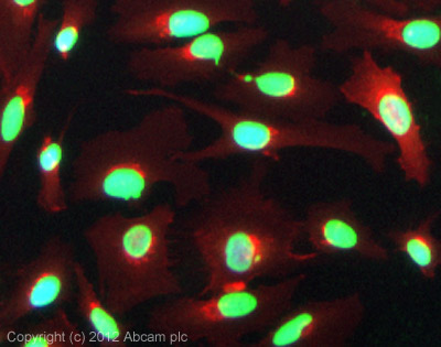 ICC/IF image of ab15584 stained HeLa cells. The cells were 100% methanol fixed (5 min) and then incubated in 1%BSA / 10% normal goat serum / 0.3M glycine in 0.1% PBS-Tween for 1h to permeabilise the cells and block non-specific protein-protein interactions. The cells were then incubated with the antibody (ab15584, 5µg/ml) overnight at +4°C. The secondary antibody (green) was ab96899, a goat anti-rabbit DyLight® 488 (IgG; H+L) used at a 1/250 dilution for 1h. Alexa Fluor® 594 WGA was used to label plasma membranes (red) at a 1/200 dilution for 1h. DAPI was used to stain the cell nuclei (blue) at a concentration of 1.43µM.
Product References
Epigenetic regulatory elements associate with specific histone modifications to - Epigenetic regulatory elements associate with specific histone modifications to
Majocchi S, Aritonovska E, Mermod N. Nucleic Acids Res. 2014 Jan;42(1):193-204.
The solution structure of the first PHD finger of autoimmune regulator in complex - The solution structure of the first PHD finger of autoimmune regulator in complex
Chignola F, Gaetani M, Rebane A, Org T, Mollica L, Zucchelli C, Spitaleri A, Mannella V, Peterson P, Musco G. Nucleic Acids Res. 2009 May;37(9):2951-61.
Arginine methylation of the histone H3 tail impedes effector binding. - Arginine methylation of the histone H3 tail impedes effector binding.
Iberg AN, Espejo A, Cheng D, Kim D, Michaud-Levesque J, Richard S, Bedford MT. J Biol Chem. 2008 Feb 8;283(6):3006-10. Epub 2007 Dec 11.
High-resolution profiling of histone methylations in the human genome. - High-resolution profiling of histone methylations in the human genome.
Barski A, Cuddapah S, Cui K, Roh TY, Schones DE, Wang Z, Wei G, Chepelev I, Zhao K. Cell. 2007 May 18;129(4):823-37.
JMJD6 is a histone arginine demethylase. - JMJD6 is a histone arginine demethylase.
Chang B, Chen Y, Zhao Y, Bruick RK. Science. 2007 Oct 19;318(5849):444-7.