Anti-Histone H2B (mono methyl K5) antibody - ChIP Grade
| Name | Anti-Histone H2B (mono methyl K5) antibody - ChIP Grade |
|---|---|
| Supplier | Abcam |
| Catalog | ab12929 |
| Prices | $380.00 |
| Sizes | 50 µg |
| Host | Rabbit |
| Clonality | Polyclonal |
| Isotype | IgG |
| Applications | IHC-P ICC/IF ICC/IF IP WB ChIP |
| Species Reactivities | Mouse, Rat, Human, Chicken, Bovine, Xenopus |
| Antigen | Synthetic peptide conjugated to KLH derived from within residues 1 - 100 of Human Histone H2B, mono methylated at K5 |
| Description | Rabbit Polyclonal |
| Gene | HIST2H2BE |
| Conjugate | Unconjugated |
| Supplier Page | Shop |
Product images
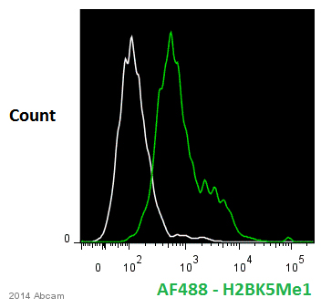 ab12929 staining Histone H2B (mono methyl K5) in human differentiated haematopoietic stem cells by Flow Cytometry. Cells were fixed with paraformaldehyde and permeabilized with permeabilization buffer. The sample was incubated with the primary antibody (1/500) for 12 hours at 4°C. An Alexa Fluor® 488-conjugated goat polyclonal anti-rabbit IgG (1/1000) was used as the secondary antibody.Gating Strategy: Isotype negative control (white).See Abreview
 Chromatin was prepared from Hela cells according to the Abcam X-ChIP protocol. Cells were fixed with formaldehyde for 10 min. The ChIP was performed with 25 µg of chromatin, 2 µg of ab12929 (blue), and 20 µl of Protein A/G sepharose beads. No antibody was added to the beads control (yellow). The immunoprecipitated DNA was quantified by real time PCR (Taqman approach). Primers and probes are located in the first kb of the transcribed region.
 All lanes : Anti-Histone H2B (mono methyl K5) antibody - ChIP Grade (ab12929) at 1 µg/mlLane 1 : Calf Thymus Histone Preparation Nuclear Lysate (ab121)Lane 2 : Calf Thymus Histone Preparation Nuclear Lysate (ab121) with Human Histone H2B (mono methyl K5) peptide (ab13211)Lane 3 : Calf Thymus Histone Preparation Nuclear Lysate (ab121) with Human Histone H2B peptide (ab13212)Lysates/proteins at 20 µg per lane.SecondaryIRDye 680 Conjugated Goat Anti-Rabbit IgG (H+L) at 1/10000 dilutionPerformed under reducing conditions.
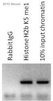 ab12929 at a 1/600 dilution for ChIP analysis of mouse dorsal skin epidermis whole tissue lysate, incubated for 15 hours at 4°C with ChIP dilution buffer. Cross-linking (X-ChIP) using 1% formaldehyde for 10 minutes.Detection step: Semiquantitative PCR.Negative control: Rabbit IgG.Cells untreated.See Abreview
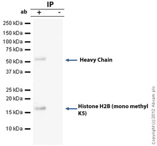 Histone H2B (mono methyl K5) was immunoprecipitated using 0.5mg Hela whole cell extract, 5µg of Rabbit polyclonal to Histone H2B (mono methyl K5) - ChIP Grade and 50µl of protein G magnetic beads (+). No antibody was added to the control (-). The antibody was incubated under agitation with Protein G beads for 10min, Hela whole cell extract lysate diluted in RIPA buffer was added to each sample and incubated for a further 10min under agitation.Proteins were eluted by addition of 40µl SDS loading buffer and incubated for 10min at 70oC; 10µl of each sample was separated on a SDS PAGE gel, transferred to a nitrocellulose membrane, blocked with 5% BSA and probed with ab12929.Secondary: Clean blot (HRP conjugate) at 1/1000 dilution.Band: 17kDa: Histone H2B (mono methyl K5).
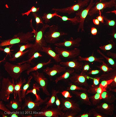 ICC/IF image of ab12929 stained HeLa cells. The cells were 100% methanol fixed (5 min) and then incubated in 1%BSA / 10% normal goat serum / 0.3M glycine in 0.1% PBS-Tween for 1h to permeabilise the cells and block non-specific protein-protein interactions. The cells were then incubated with the antibody ab12929 at 1µg/ml overnight at +4°C. The secondary antibody (green) was a goat anti-rabbit DyLight® 488 (ab96899) IgG (H+L) used at a 1/1000 dilution for 1h. Alexa Fluor® 594 WGA was used to label plasma membranes (red) at a 1/200 dilution for 1h. DAPI was used to stain the cell nuclei (blue) at a concentration of 1.43µM.
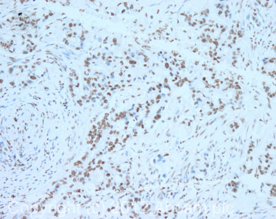 IHC image of Histone H2B (mono methyl K5) staining in human pancreatic adenocarcinoma formalin fixed paraffin embedded tissue section, performed on a Leica BondTM system using the standard protocol F. The section was pre-treated using heat mediated antigen retrieval with sodium citrate buffer (pH6, epitope retrieval solution 1) for 20 mins. The section was then incubated with ab12929, 0.1µg/ml, for 15 mins at room temperature and detected using an HRP conjugated compact polymer system. DAB was used as the chromogen. The section was then counterstained with haematoxylin and mounted with DPX. For other IHC staining systems (automated and non-automated) customers should optimize variable parameters such as antigen retrieval conditions, primary antibody concentration and antibody incubation times.
Product References
Epigenetic regulatory elements associate with specific histone modifications to - Epigenetic regulatory elements associate with specific histone modifications to
Majocchi S, Aritonovska E, Mermod N. Nucleic Acids Res. 2014 Jan;42(1):193-204.
The activity-dependent histone variant H2BE modulates the life span of olfactory - The activity-dependent histone variant H2BE modulates the life span of olfactory
Santoro SW, Dulac C. Elife. 2012 Dec 13;1:e00070.
Combinatorial patterns of histone acetylations and methylations in the human - Combinatorial patterns of histone acetylations and methylations in the human
Wang Z, Zang C, Rosenfeld JA, Schones DE, Barski A, Cuddapah S, Cui K, Roh TY, Peng W, Zhang MQ, Zhao K. Nat Genet. 2008 Jul;40(7):897-903.
High-resolution profiling of histone methylations in the human genome. - High-resolution profiling of histone methylations in the human genome.
Barski A, Cuddapah S, Cui K, Roh TY, Schones DE, Wang Z, Wei G, Chepelev I, Zhao K. Cell. 2007 May 18;129(4):823-37.