![Anti-Histone H2B (crotonyl K5) antibody [EPR17483] (ab177396) at 1/1000 dilution + HeLa (Human epithelial cells from cervix adenocarcinoma) cell lysate at 20 µgSecondaryGoat Anti-Rabbit IgG, (H+L), Peroxidase conjugated at 1/1000 dilution](http://www.bioprodhub.com/system/product_images/ab_products/2/sub_3/1347_ab177396-236868-177396.jpg)
Anti-Histone H2B (crotonyl K5) antibody [EPR17483] (ab177396) at 1/1000 dilution + HeLa (Human epithelial cells from cervix adenocarcinoma) cell lysate at 20 µgSecondaryGoat Anti-Rabbit IgG, (H+L), Peroxidase conjugated at 1/1000 dilution
![Anti-Histone H2B (crotonyl K5) antibody [EPR17483] (ab177396) at 1/500 dilution + NIH/3T3 (Mouse embyro fibroblast cells) cell lysate at 10 µgSecondaryGoat Anti-Rabbit IgG, (H+L), Peroxidase conjugated at 1/1000 dilution](http://www.bioprodhub.com/system/product_images/ab_products/2/sub_3/1348_ab177396-236867-1773962.jpg)
Anti-Histone H2B (crotonyl K5) antibody [EPR17483] (ab177396) at 1/500 dilution + NIH/3T3 (Mouse embyro fibroblast cells) cell lysate at 10 µgSecondaryGoat Anti-Rabbit IgG, (H+L), Peroxidase conjugated at 1/1000 dilution
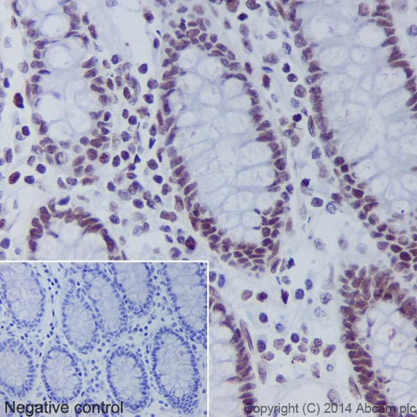
Immunohistochemical analysis of paraffin-embedded Human colon tissue labeling Histone H2B (crotonyl K5) using ab177396 at 1/500 dilution. A Goat Anti-Rabbit IgG H&L (HRP) (ab97051) was used as secondary at 1/500 dilution. Counter stained with Hematoxylin.Inset image: negative control obtained using PBS instead of ab177396.Note: Nucleus staining on glandular epithelium of Human colon tissue was observed.
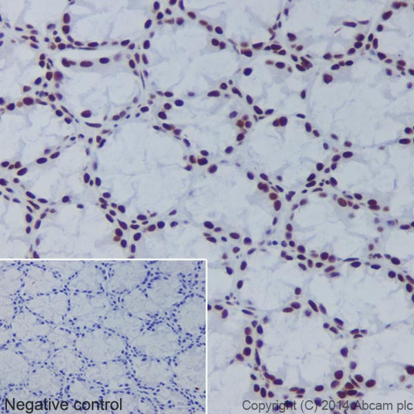
Immunohistochemical analysis of paraffin-embedded Mouse colon tissue labeling Histone H2B (crotonyl K5) using ab177396 at 1/500 dilution. A Goat Anti-Rabbit IgG H&L (HRP) (ab97051) was used as secondary at 1/500 dilution. Counter stained with Hematoxylin.Inset image: negative control obtained using PBS instead of ab177396.Note: Nucleus staining on glandular epithelium of Mouse colon tissue was observed.
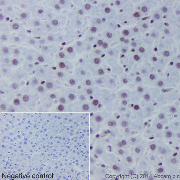
Immunohistochemical analysis of paraffin-embedded Rat liver tissue labeling Histone H2B (crotonyl K5) using ab177396 at 1/500 dilution. A Goat Anti-Rabbit IgG H&L (HRP) (ab97051) was used as secondary at 1/500 dilution. Counter stained with Hematoxylin.Inset image: negative control obtained using PBS instead of ab177396.Note: Nucleus staining on Rat liver tissue was observed.
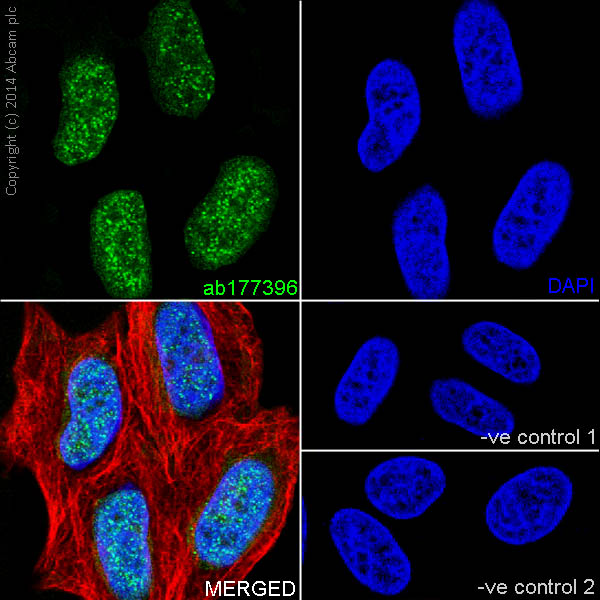
Immunofluorescent analysis of 4% paraformaldehyde-fixed, 0.1% Triton X-100 permeabilized HeLa (Human epithelial cells from cervix adenocarcinoma) cells labeling Histone H2B (crotonyl K5) with ab177396 at 1/250 dilution, followed by Goat anti-rabbit IgG (Alexa Fluor® 488) (ab150077) secondary antibody at 1/500 dilution (green). Confocal image showing nuclear staining on HeLa cell line. The nuclear counter stain is DAPI (blue). Tubulin is detected with ab7291 (anti-Tubulin mouse mAb) at 1/1000 dilution and ab150120 (AlexaFluor®594 Goat anti-Mouse secondary) at 1/500 dilution (red).The negative controls are as follows:1. ab177396 at 1/250 dilution followed by ab150120 (AlexaFluor®594 Goat anti-Mouse secondary) at 1/500 dilution.2. ab7291 (anti-Tubulin mouse mAb) at 1/1000 dilution followed by ab150077 (Alexa Fluor®488 Goat Anti-Rabbit IgG H&L) at 1/500 dilution.