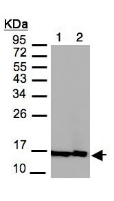
All lanes : Anti-Histone H2A.Z antibody - ChIP Grade (ab97966) at 1/3000 dilutionLane 1 : MOLT4 whole cell lysateLane 2 : Raji whole cell lysateLysates/proteins at 30 µg per lane.
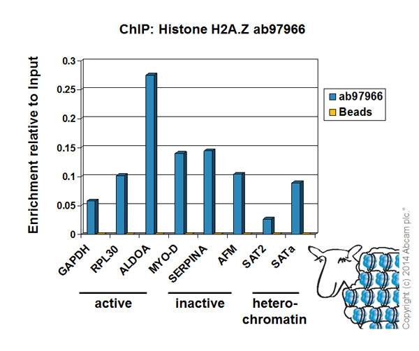
Chromatin was prepared from HeLa cells according to the Abcam X-ChIP protocol. Cells were fixed with formaldehyde for 10 minutes. The ChIP was performed with 25µg of chromatin, 2µg of ab97966 (blue), and 20µl of Protein A/G sepharose beads. No antibody was added to the beads control (yellow). The immunoprecipitated DNA was quantified by real time PCR (Taqman approach for active and inactive loci, Sybr green approach for heterochromatic loci). Primers and probes are located in the first kb of the transcribed region.
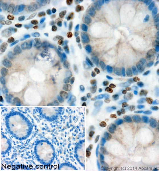
IHC image of ab97966 staining Histone H3 (phospho T45) in human colon formalin fixed paraffin embedded tissue sections*, performed on a Leica Bond. The section was pre-treated using heat mediated antigen retrieval with sodium citrate buffer (pH6, epitope retrieval solution 1) for 20 mins. The section was then incubated with ab97966, 1µg/ml, for 15 mins at room temperature and detected using an HRP conjugated compact polymer system. DAB was used as the chromogen. The section was then counterstained with haematoxylin and mounted with DPX. No primary antibody was used in the negative control (shown on the inset).For other IHC staining systems (automated and non-automated) customers should optimize variable parameters such as antigen retrieval conditions, primary antibody concentration and antibody incubation times.*Tissue obtained from the Human Research Tissue Bank, supported by the NIHR Cambridge Biomedical Research Centre
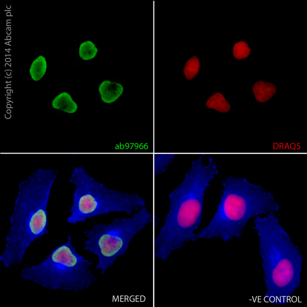
ab97966 staining Histone H2A.Z in HeLa cells. The cells were fixed with 100% methanol (5min) and then blocked in 1% BSA/10% normal goat serum/0.3M glycine in 0.1%PBS-Tween for 1h. The cells were then incubated with ab97966 at 0.5μg/ml overnight at +4°C, followed by a further incubation at room temperature for 1h with an AlexaFluor®488 Goat anti-Rabbit secondary (ab150077) at 2 μg/ml (shown in green). AlexaFluor®350 WGA was used at a 1/200 dilution and incubated for 1h with the cells, to label plasma membranes (shown in blue). Nuclear DNA was labelled in red with 1.25 μM DRAQ5™ (ab108410), which was added to the secondary antibody mixture. A secondary only negative control is displayed, which indicates that the Histone H2A.Z staining observed is due to primary antibody specificity and not to unspecific binding of the secondary antibody to the cells.