Anti-Histone H2A antibody
| Name | Anti-Histone H2A antibody |
|---|---|
| Supplier | Abcam |
| Catalog | ab93460 |
| Prices | $359.00 |
| Sizes | 100 µg |
| Host | Rabbit |
| Clonality | Polyclonal |
| Isotype | IgG |
| Applications | WB FC IHC-P |
| Species Reactivities | Human |
| Antigen | Synthetic peptide corresponding to Human Histone H2A (N terminal) conjugated to Keyhole Limpet Haemocyanin (KLH) |
| Description | Rabbit Polyclonal |
| Gene | HIST1H2AE |
| Conjugate | Unconjugated |
| Supplier Page | Shop |
Product images
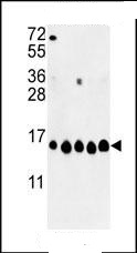 All lanes : Anti-Histone H2A antibody (ab93460) at 1/1000 dilutionLane 1 : A2058Lane 2 : A375Lane 3 : K562Lane 4 : HeLaLane 5 : MCF-7Lysates/proteins at 35 µg per lane.
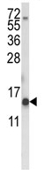 Anti-Histone H2A antibody (ab93460) at 1/100 dilution + K562 cell line lysates at 35 µg
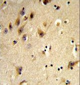 Formalin-fixed and paraffin-embedded human brain tissue reacted with AB93460 at 1/50 dilution, which was peroxidase-conjugated to the secondary antibody, followed by DAB staining.
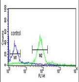 ab93460 cytometric analysis of MCF-7 cells (right histogram) compared to a negative control cell (left histogram).FITC-conjugatedgoat-anti-rabbit secondary antibodies were used for the analysis.