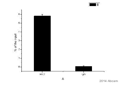
ChIP analysis using ab181973 binding Histone H1.2 in mouse primary hepatocytes. Cells were cross-linked for 10 minutes with 1% formaldehyde. Samples were incubated with the primary antibody (1/100) for 20 hours at 4°C. Protein binding was detected using real-time PCR.Negative Control: IgG.See Abreview
![All lanes : Anti-Histone H1.2 antibody [EPR12690] (ab181973) at 1/10000 dilutionLane 1 : HeLa cell lysateLane 2 : A375 cell lysateLane 3 : 293 cell lysateLysates/proteins at 20 µg per lane.SecondaryGoat Anti-Rabbit IgG, (H+L), Peroxidase conjugate at 1/1000 dilution](http://www.bioprodhub.com/system/product_images/ab_products/2/sub_3/946_ab181973-212231-ab181973WB.jpg)
All lanes : Anti-Histone H1.2 antibody [EPR12690] (ab181973) at 1/10000 dilutionLane 1 : HeLa cell lysateLane 2 : A375 cell lysateLane 3 : 293 cell lysateLysates/proteins at 20 µg per lane.SecondaryGoat Anti-Rabbit IgG, (H+L), Peroxidase conjugate at 1/1000 dilution
![Anti-Histone H1.2 antibody [EPR12690] (ab181973) at 1/2000 dilution + MCF7 cell lysate at 20 µgSecondaryGoat Anti-Rabbit IgG, (H+L), Peroxidase conjugate at 1/1000 dilution](http://www.bioprodhub.com/system/product_images/ab_products/2/sub_3/947_ab181973-212229-ab181973WBb.jpg)
Anti-Histone H1.2 antibody [EPR12690] (ab181973) at 1/2000 dilution + MCF7 cell lysate at 20 µgSecondaryGoat Anti-Rabbit IgG, (H+L), Peroxidase conjugate at 1/1000 dilution
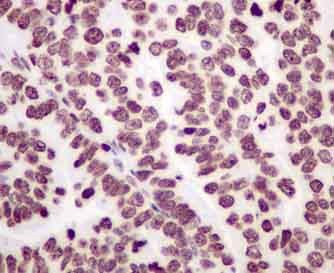
Immunohistochemical analysis of paraffin-embedded Human ovarian carcinoma tissue labeling Histone H1.2 with ab181973 at 1/100 dilution, followed by prediluted ImmunoHistoprobe (Ready to use) HRP Polymer for Rabbit IgG. Counter stained with Hematoxylin.
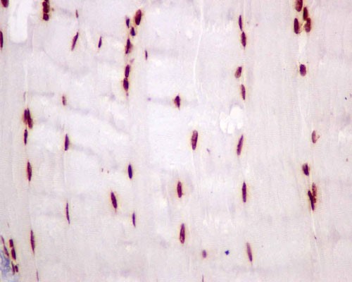
Immunohistochemical analysis of paraffin-embedded Mouse muscle tissue labeling Histone H1.2 with ab181973 at 1/100 dilution, followed by prediluted ImmunoHistoprobe (Ready to use) HRP Polymer for Rabbit IgG. Counter stained with Hematoxylin.
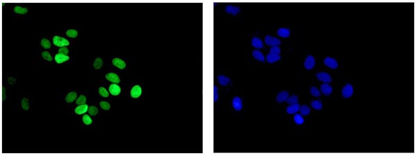
Immunofluorescent analysis of 4% paraformaldehyde-fixed MCF7 cells labeling Histone H1.2 with ab181973 at 1/250 dilution, followed by Goat anti rabbit IgG (Dylight 488) at 1/200 dilution (green), Dapi staining (blue).