![All lanes : Anti-HIF1 beta antibody [1A1] (ab123853) at 1/2000 dilutionLane 1 : HEK293T cells transfected with pCMV6-ENTRY control cDNALane 2 : HEK293T cells transfected with pCMV6-ENTRY HIF1 beta cDNALysates/proteins at 5 µg per lane.](http://www.bioprodhub.com/system/product_images/ab_products/2/sub_3/632_HIF1-beta-Primary-antibodies-ab123853-1.jpg)
All lanes : Anti-HIF1 beta antibody [1A1] (ab123853) at 1/2000 dilutionLane 1 : HEK293T cells transfected with pCMV6-ENTRY control cDNALane 2 : HEK293T cells transfected with pCMV6-ENTRY HIF1 beta cDNALysates/proteins at 5 µg per lane.
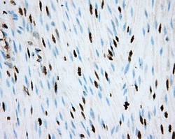
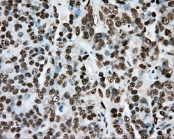
ab123853, at 1/50 dilution, staining HIF1 beta in paraffin-embedded Human colon tissue by Immunohistochemistry.
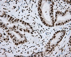
ab123853, at 1/50 dilution, staining HIF1 beta in paraffin-embedded Human colon adenocarcinoma tissue by Immunohistochemistry.
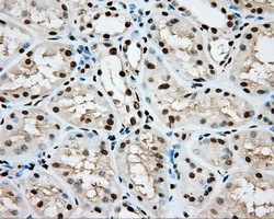
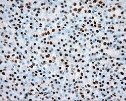
ab123853, at 1/50 dilution, staining HIF1 beta in paraffin-embedded Human kidney tissue by Immunohistochemistry.
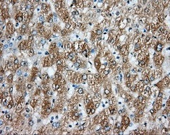
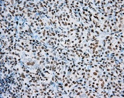
ab123853, at 1/50 dilution, staining HIF1 beta in paraffin-embedded Human liver tissue by Immunohistochemistry.
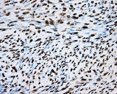
ab123853, at 1/50 dilution, staining HIF1 beta in paraffin-embedded Human ovary adenocarcinoma tissue by Immunohistochemistry.
ab123853, at 1/50 dilution, staining HIF1 beta in paraffin-embedded Human pancreas tissue by Immunohistochemistry.
ab123853, at 1/50 dilution, staining HIF1 beta in paraffin-embeddedHuman thyroid carcinoma tissue by Immunohistochemistry.
ab123853, at 1/50 dilution, staining HIF1 beta in paraffin-embedded Human endometrium tissue by Immunohistochemistry.
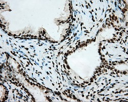
ab123853, at 1/50 dilution, staining HIF1 beta in paraffin-embedded Human prostate tissue by Immunohistochemistry.
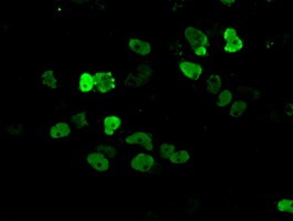
ab123853, at 1/100 dilution, staining HIF1 beta in COS7 cells transiently transfected by pCMV6-ENTRY HIF1 beta by Immunofluorescence.
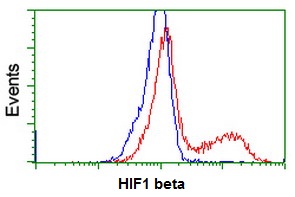
ab123853, at 1/100 dilution, staining HIF1 beta in HEK293T cells transfected with either pCMV6-ENTRY HIF1-beta (Red) or empty vector control (Blue) analysed by flow cytometry.
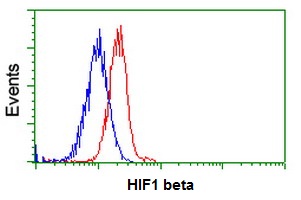
ab123853, at 1/100 dilution staining HIF1 beta in Jurkat cells by Flow cytometry (Red) compared to a nonspecific negative control antibody (Blue).