Anti-Hes1 antibody [EPR4226]
| Name | Anti-Hes1 antibody [EPR4226] |
|---|---|
| Supplier | Abcam |
| Catalog | ab108937 |
| Prices | $391.00 |
| Sizes | 100 µl |
| Host | Rabbit |
| Clonality | Monoclonal |
| Isotype | IgG |
| Clone | EPR4226 |
| Applications | ICC/IF ICC/IF WB IHC-P FC |
| Species Reactivities | Mouse, Rat, Human |
| Antigen | Synthetic peptide (the amino acid sequence is considered to be commercially sensitive) corresponding to Human Hes1 aa 200-300 |
| Description | Rabbit Monoclonal |
| Gene | HES1 |
| Conjugate | Unconjugated |
| Supplier Page | Shop |
Product images
![Anti-Hes1 antibody [EPR4226] (ab108937) at 1/1000 dilution (purified) + SH-SY5Y cell lysate at 10 µgSecondaryHRP goat anti-rabbit (H+L) at 1/1000 dilution](http://www.bioprodhub.com/system/product_images/ab_products/2/sub_3/91_ab108937-239403-108937-WB-2.jpg) Anti-Hes1 antibody [EPR4226] (ab108937) at 1/1000 dilution (purified) + SH-SY5Y cell lysate at 10 µgSecondaryHRP goat anti-rabbit (H+L) at 1/1000 dilution
Anti-Hes1 antibody [EPR4226] (ab108937) at 1/1000 dilution (purified) + SH-SY5Y cell lysate at 10 µgSecondaryHRP goat anti-rabbit (H+L) at 1/1000 dilution
 Immunofluorescent staining of SH-SY5Y cells (fixed with 4% PFA and permeablized with TritonX 100) with purified ab108937 at a dilution of 1/100. An Alexa Fluor® 555 goat anti-rabbit antibody was used as the secondary at a dilution of 1/200. The panel on the right shows the DAPI counter-staining.
 Overlay histogram showing SH-SY5Y cells fixed in 2% PFA and stained with purified ab108937 at a dilution of 1 in 30 (pink line). The secondary antibody used was FITC goat anti-rabbit at a dilution of 1 in 50. Rabbit monoclonal IgG was used as an isotype control.
![All lanes : Anti-Hes1 antibody [EPR4226] (ab108937) at 1/1000 dilution (unpurified)Lane 1 : Fetal brain lysateLane 2 : PC12 cell lysateLane 3 : SH-SY5Y cell lysateLysates/proteins at 10 µg per lane.SecondaryHRP-labelled goat anti-rabbit at 1/2000 dilution](http://www.bioprodhub.com/system/product_images/ab_products/2/sub_3/95_Hes1-Primary-antibodies-ab108937-1.JPG) All lanes : Anti-Hes1 antibody [EPR4226] (ab108937) at 1/1000 dilution (unpurified)Lane 1 : Fetal brain lysateLane 2 : PC12 cell lysateLane 3 : SH-SY5Y cell lysateLysates/proteins at 10 µg per lane.SecondaryHRP-labelled goat anti-rabbit at 1/2000 dilution
All lanes : Anti-Hes1 antibody [EPR4226] (ab108937) at 1/1000 dilution (unpurified)Lane 1 : Fetal brain lysateLane 2 : PC12 cell lysateLane 3 : SH-SY5Y cell lysateLysates/proteins at 10 µg per lane.SecondaryHRP-labelled goat anti-rabbit at 1/2000 dilution
![Anti-Hes1 antibody [EPR4226] (ab108937) at 1/1000 dilution (unpurified) + SH-SY5Y cell lysate at 10 µgSecondaryHRP goat anti-rabbit (H+L) at 1/1000 dilution](http://www.bioprodhub.com/system/product_images/ab_products/2/sub_3/96_ab108937-239402-108937-WB-1.jpg) Anti-Hes1 antibody [EPR4226] (ab108937) at 1/1000 dilution (unpurified) + SH-SY5Y cell lysate at 10 µgSecondaryHRP goat anti-rabbit (H+L) at 1/1000 dilution
Anti-Hes1 antibody [EPR4226] (ab108937) at 1/1000 dilution (unpurified) + SH-SY5Y cell lysate at 10 µgSecondaryHRP goat anti-rabbit (H+L) at 1/1000 dilution
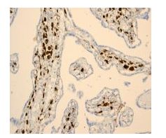 Immunohistochemical staining of Hes1 in paraffin-embedded human placenta tissue with unpurified ab108937 at 1/250 dilution.
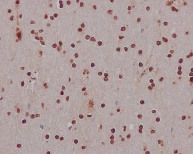
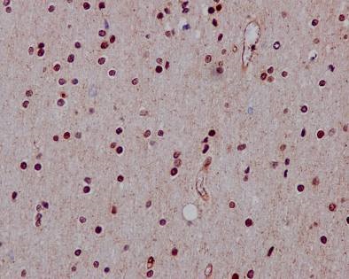 Immunohistochemical staining of paraffin embedded human brain with unpurified ab108937 at a working dilution of 1 in 90. The secondary antibody used is a HRP polymer for rabbit IgG. The sample is counter-stained with hematoxylin. Antigen retrieval was perfomed using Tris-EDTA buffer, pH 9.0.
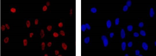
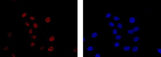 Immunofluorescent staining of SH-SY5Y cells (fixed with 4% PFA and permeablized with TritonX 100) with unpurified ab108937 at a dilution of 1/40. An Alexa Fluor® 555 goat anti-rabbit antibody was used as the secondary at a dilution of 1/200. The panel on the right shows the DAPI counter-staining.
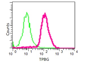
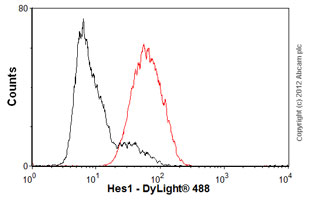 Overlay histogram showing SH-SY5Y cells stained with unpurified ab108937 (red line). The cells were fixed with 80% methanol (5 min) and then permeabilized with 0.1% PBS-Tween for 20 min. The cells were then incubated in 1x PBS / 10% normal goat serum / 0.3M glycine to block non-specific protein-protein interactions. The cells were then incubated with the antibody (ab108937, 1/100 dilution) for 30 min at 22ºC. The secondary antibody used was DyLight® 488 goat anti-rabbit IgG (H+L) (ab96899) at 1/500 dilution for 30 min at 22ºC. Isotype control antibody (black line) was rabbit IgG (monoclonal) (1µg/1x106 cells) used under the same conditions. Acquisition of >5,000 events was performed.
 Overlay histogram showing SH-SY5Y cells fixed in 2% PFA and stained with unpurified ab108937 at a dilution of 1 in 30 (pink line). The secondary antibody used was FITC goat anti-rabbit at a dilution of 1 in 20. Rabbit monoclonal IgG was used as an isotype control.
Product References
Alterations in Notch signalling in skeletal muscles from mdx and dko dystrophic - Alterations in Notch signalling in skeletal muscles from mdx and dko dystrophic
Church JE, Trieu J, Chee A, Naim T, Gehrig SM, Lamon S, Angelini C, Russell AP, Lynch GS. Exp Physiol. 2014 Apr;99(4):675-87.
HES1, a target of Notch signaling, is elevated in canine osteosarcoma, but - HES1, a target of Notch signaling, is elevated in canine osteosarcoma, but
Dailey DD, Anfinsen KP, Pfaff LE, Ehrhart EJ, Charles JB, Bonsdorff TB, Thamm DH, Powers BE, Jonasdottir TJ, Duval DL. BMC Vet Res. 2013 Jul 1;9:130.
Reduction of NOTCH1 expression pertains to maturation abnormalities of - Reduction of NOTCH1 expression pertains to maturation abnormalities of
Sakamoto K, Fujii T, Kawachi H, Miki Y, Omura K, Morita K, Kayamori K, Katsube K, Yamaguchi A. Lab Invest. 2012 May;92(5):688-702.