Anti-HADHA antibody
| Name | Anti-HADHA antibody |
|---|---|
| Supplier | Abcam |
| Catalog | ab137831 |
| Prices | $384.00 |
| Sizes | 100 µl |
| Host | Rabbit |
| Clonality | Polyclonal |
| Isotype | IgG |
| Applications | WB IHC-P ICC/IF ICC/IF |
| Species Reactivities | Mouse, Rat, Human |
| Antigen | Recombinant fragment corresponding to a region within amino acids 152-454 of Human HADHA (UniProt ID: P40939) |
| Description | Rabbit Polyclonal |
| Gene | HADHA |
| Conjugate | Unconjugated |
| Supplier Page | Shop |
Product images
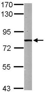
 All lanes : Anti-HADHA antibody (ab137831) at 1/1000 dilutionLane 1 : 293T whole cell lysateLane 2 : A431 whole cell lysateLysates/proteins at 30 µg per lane.
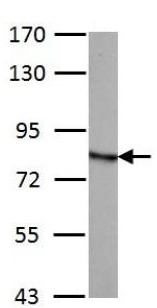
 Anti-HADHA antibody (ab137831) at 1/1000 dilution + Mouse liver whole cell lysate at 50 µg
 Anti-HADHA antibody (ab137831) at 1/500 dilution + PC12 whole cell lysate at 30 µg
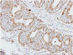 Immunohistochemical analysis of paraffin embedded colon carcinoma tissue, labelling HADHA using ab137831 at 1/250 dilution.
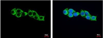 Immunofluorescence analysis of methanol fixed (100% methanol at -20 °C for 5 minutes) HepG2 cells, labelling HADHA using ab137831 at 1/500 dilution. Right panel (blue) shows Hoechst 33343 staining.