Anti-GRP78 BiP antibody
| Name | Anti-GRP78 BiP antibody |
|---|---|
| Supplier | Abcam |
| Catalog | ab21685 |
| Prices | $401.00 |
| Sizes | 100 µg |
| Host | Rabbit |
| Clonality | Polyclonal |
| Isotype | IgG |
| Applications | IHC-F IP ICC/IF ICC/IF WB Electron microscopy IHC-P IHC-F |
| Species Reactivities | Mouse, Rat, Dog, Human, Pig, Monkey, Hamster |
| Antigen | Synthetic peptide conjugated to KLH derived from within residues 600 to the C-terminus of Mouse GRP78 BiP |
| Blocking Peptide | Mouse GRP78 BiP peptide |
| Description | Rabbit Polyclonal |
| Gene | Hspa5 |
| Conjugate | Unconjugated |
| Supplier Page | Shop |
Product images
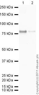
 All lanes : Anti-GRP78 BiP antibody (ab21685) at 1 µg/mlLane 1 : CHO-K1 whole cell lysate at 20 µgLane 2 : Liver (Mouse) Tissue Lysate at 20 µgLane 3 : Rat liver whole cell lysate at 20 µgLane 4 : HeLa whole cell lysate at 20 µgLane 5 : CHO-K1 whole cell lysate at 20 µg/ml with Mouse GRP78 BiP peptide (ab22410) at 1 µgLane 6 : Liver (Mouse) Tissue Lysate at 20 µg with Mouse GRP78 BiP peptide (ab22410) at 1 µg/mlLane 7 : Rat liver whole cell lysate at 20 µg with Mouse GRP78 BiP peptide (ab22410) at 1 µg/mlLane 8 : HeLa whole cell lysate at 20 µg with Mouse GRP78 BiP peptide (ab22410) at 1 µg/mlSecondaryGoat anti Rabbit IgG at 1/10000 dilutionPerformed under reducing conditions.
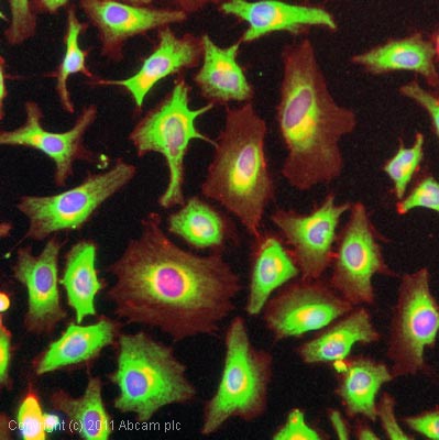
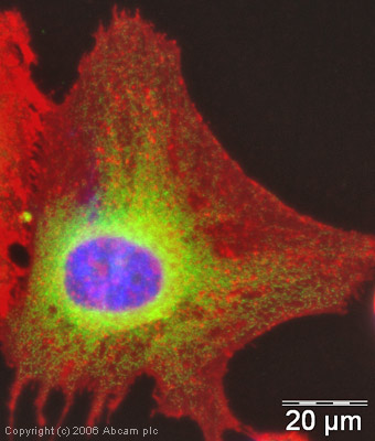 ICC/IF image of ab21685 stained human HeLa cells. The cells were methanol fixed (5 min) and incubated with the antibody (ab21685, 1µg/ml) for 1h at room temperature. The secondary antibody (green) was Alexa Fluor® 488 goat anti-rabbit IgG (H+L) used at a 1/1000 dilution for 1h. Image-iTTM FX Signal Enhancer was used as the primary blocking agent, 5% BSA (in TBS-T) was used for all other blocking steps. DAPI was used to stain the cell nuclei (blue). Alexa Fluor® 594 WGA was used to label plasma membranes (red).
 ICC/IF image of ab21685 stained human HeLa cells. The cells were methanol fixed (5 min) and incubated with the antibody (ab21685, 1µg/ml) for 1h at room temperature. The secondary antibody (green) was Alexa Fluor® 488 goat anti-rabbit IgG (H+L) used at a 1/1000 dilution for 1h. Image-iTTM FX Signal Enhancer was used as the primary blocking agent, 5% BSA (in TBS-T) was used for all other blocking steps. DAPI was used to stain the cell nuclei (blue). Alexa Fluor® 594 WGA was used to label plasma membranes (red).
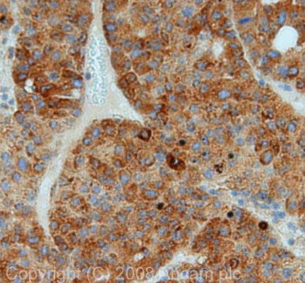 IHC image of GRP78 BiP staining in human liver carcinoma FFPE section, performed on a BondTM system using the standard protocol F. The section was pre-treated using heat mediated antigen retrieval with sodium citrate buffer (pH6, epitope retrieval solution 1) for 20 mins. The section was then incubated with ab21685, 1µg/ml, for 8 mins at room temperature and detected using an HRP conjugated compact polymer system. DAB was used as the chromogen. The section was then counterstained with haematoxylin and mounted with DPX.
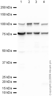 All lanes : Anti-GRP78 BiP antibody (ab21685) at 1 µg/mlLane 1 : CHO-K1 cell lysate Whole Cell LysateLane 2 : Liver (Mouse) Tissue Lysate at 10 µgLane 3 : Liver (Rat) Tissue Lysate at 10 µgLane 4 : HeLa (Human epithelial carcinoma cell line) Whole Cell Lysate at 10 µgSecondaryGoat polyclonal to Rabbit IgG - H&L - Pre-Adsorbed (HRP) at 1/3000 dilutiondeveloped using the ECL techniquePerformed under reducing conditions.
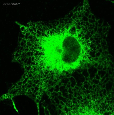 ab21685 staining GRP78 BiP in African Green Monkey COS-7 cells by ICC/IF (Immunocytochemistry/immunofluorescence). Cells were fixed with paraformaldehyde, permeabilized with 0.5% Triton X-100 and blocked with 3% BSA for 1 hour at 23°C. Samples were incubated with primary antibody (1/1000 in PBS-BSA) for 1 hour at 23°C. An Alexa Fluor® 488-conjugated Goat anti-rabbit IgG polyclonal (1/1000) was used as the secondary antibody.See Abreview
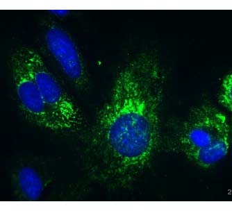 ab21685 at a 1/500 dilution staining GRP78 BiP in mouse retinal pigment epithelium primary cells by Immunocytochemistry/ Immunofluorescence, incubated for 16 hours at 4°C. PFA fixed. Blocked with 5% serum for 20 minutes at 25°C. Secondary used at a 1/500 dilution polyclonal Goat anti-rabbit conjugated to Alexa Fluor 488 (green). Nuclei were counterstained with DAPI (blue).See Abreview
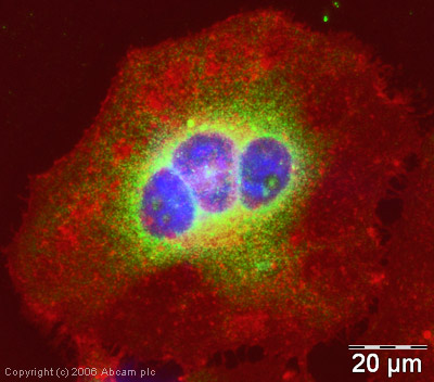
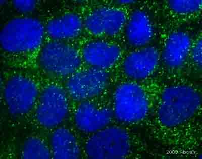 ab21685 at a 1/500 dilution staining GRP78 BiP in Dog MDCK II cells by Immunocytochemistry/ Immunofluorescence, incubated for 16 hours at 4°C. PFA fixed. Blocked with 5% serum for 20 minutes at 25°C. Secondary used at a 1/500 dilution polyclonal Goat anti-rabbit conjugated to Alexa Fluor 488 (green). Nuclei were counterstained with DAPI (blue).See Abreview
 ICC/IF image of ab21685 stained Hela cells. The cells were 100% methanol fixed (5 min) and then incubated in 1%BSA / 10% normal goat serum / 0.3M glycine in 0.1% PBS-Tween for 1h to permeabilise the cells and block non-specific protein-protein interactions. The cells were then incubated with the antibody (ab21685, 1µg/ml) overnight at +4°C. The secondary antibody (green) was DyLight® 488 goat anti-rabbit IgG - H&L, pre-adsorbed (ab96899) used at a 1/250 dilution for 1h. Alexa Fluor® 594 WGA was used to label plasma membranes (red) at a 1/200 dilution for 1h. DAPI was used to stain the cell nuclei (blue) at a concentration of 1.43µM.
 developed using the ECL techniquePerformed under reducing conditions.
 developed using the ECL techniquePerformed under reducing conditions.
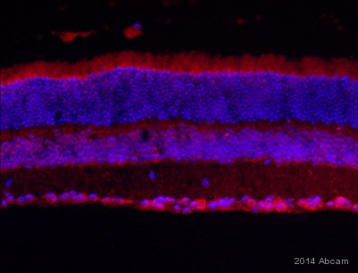 ab21685 staining GRP78 BiP in Mouse retina tissue sections by Immunohistochemistry (PFA perfusion fixed frozen sections). Tissue samples were fixed by perfusion with paraformaldehyde, permeablized with Triton X-100, blocked with 1% BSA for 1 hour at 25°C. The sample was incubated with primary antibody (1/400 in PBST + BSA + goat serum) at 4°C for 17 hours. A DyLight® 650-conjugated goat anti-rabbit polyclonal (1/600) was used as the secondary antibody.See Abreview
Product References
Anti-cancer effects of REIC/Dkk-3-encoding adenoviral vector for the treatment of - Anti-cancer effects of REIC/Dkk-3-encoding adenoviral vector for the treatment of
Shien K, Tanaka N, Watanabe M, Soh J, Sakaguchi M, Matsuo K, Yamamoto H, Furukawa M, Asano H, Tsukuda K, Nasu Y, Huh NH, Miyoshi S, Kumon H, Toyooka S. PLoS One. 2014 Feb 3;9(2):e87900.
Generation of human ER chaperone BiP in yeast Saccharomyces cerevisiae. - Generation of human ER chaperone BiP in yeast Saccharomyces cerevisiae.
Ciplys E, Aucynaite A, Slibinskas R. Microb Cell Fact. 2014 Feb 11;13:22.
The i-AAA protease YME1L and OMA1 cleave OPA1 to balance mitochondrial fusion and - The i-AAA protease YME1L and OMA1 cleave OPA1 to balance mitochondrial fusion and
Anand R, Wai T, Baker MJ, Kladt N, Schauss AC, Rugarli E, Langer T. J Cell Biol. 2014 Mar 17;204(6):919-29.
No amelioration of uromodulin maturation and trafficking defect by sodium - No amelioration of uromodulin maturation and trafficking defect by sodium
Kemter E, Sklenak S, Rathkolb B, Hrabe de Angelis M, Wolf E, Aigner B, Wanke R. J Biol Chem. 2014 Apr 11;289(15):10715-26.
HKH40A downregulates GRP78/BiP expression in cancer cells. - HKH40A downregulates GRP78/BiP expression in cancer cells.
Kosakowska-Cholody T, Lin J, Srideshikan SM, Scheffer L, Tarasova NI, Acharya JK. Cell Death Dis. 2014 May 22;5:e1240.
.
Tapasin-related protein TAPBPR is an additional component of the MHC class I - Tapasin-related protein TAPBPR is an additional component of the MHC class I
Boyle LH, Hermann C, Boname JM, Porter KM, Patel PA, Burr ML, Duncan LM, Harbour ME, Rhodes DA, Skjodt K, Lehner PJ, Trowsdale J. Proc Natl Acad Sci U S A. 2013 Feb 26;110(9):3465-70. doi:
Role of C-terminal membrane-proximal basic residues in cell surface trafficking - Role of C-terminal membrane-proximal basic residues in cell surface trafficking
Okamoto Y, Bernstein JD, Shikano S. J Biol Chem. 2013 Mar 29;288(13):9189-99.
Type of uromodulin mutation and allelic status influence onset and severity of - Type of uromodulin mutation and allelic status influence onset and severity of
Kemter E, Prueckl P, Sklenak S, Rathkolb B, Habermann FA, Hans W, Gailus-Durner V, Fuchs H, Hrabe de Angelis M, Wolf E, Aigner B, Wanke R. Hum Mol Genet. 2013 Oct 15;22(20):4148-63.
New role of signal peptide peptidase to liberate C-terminal peptides for MHC - New role of signal peptide peptidase to liberate C-terminal peptides for MHC
Oliveira CC, Querido B, Sluijter M, de Groot AF, van der Zee R, Rabelink MJ, Hoeben RC, Ossendorp F, van der Burg SH, van Hall T. J Immunol. 2013 Oct 15;191(8):4020-8.