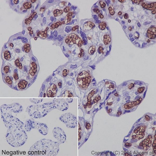
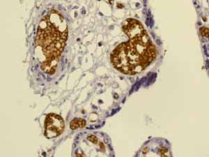
ab129024 staining Glycophorin A in Human placenta tissue sections by Immunohistochemistry (IHC-P - paraformaldehyde-fixed, paraffin-embedded sections). Tissue was fixed and paraffin-embedded, antigen retrieval was by heat mediation in Tris/EDTA buffer pH9. Samples were incubated with primary antibody (1/2500). ab97051(1/500) HRP-conjugated goat anti-rabbit IgG(H&L) was used as the secondary antibody. Tissue counterstained with Hematoxylin. PBS was used in the negative control rather than the Primary antibody.
![Anti-Glycophorin A antibody [EPR8200] (ab129024) at 1/1000 dilution + Human fetal kidney at 10 µgSecondaryGoat Anti-Rabbit IgG, (H+L), HRP- conjugated at 1/1000 dilution](http://www.bioprodhub.com/system/product_images/ab_products/2/sub_2/25822_ab129024-239920-129024-WB2.jpg)
Anti-Glycophorin A antibody [EPR8200] (ab129024) at 1/1000 dilution + Human fetal kidney at 10 µgSecondaryGoat Anti-Rabbit IgG, (H+L), HRP- conjugated at 1/1000 dilution
![Anti-Glycophorin A antibody [EPR8200] (ab129024) at 1/2000 dilution + Human fetal liver lysate at 20 µgSecondaryGoat Anti-Rabbit IgG, (H+L), HRP- conjugated at 1/1000 dilution](http://www.bioprodhub.com/system/product_images/ab_products/2/sub_2/25823_ab129024-239919-129024-WB1.jpg)
Anti-Glycophorin A antibody [EPR8200] (ab129024) at 1/2000 dilution + Human fetal liver lysate at 20 µgSecondaryGoat Anti-Rabbit IgG, (H+L), HRP- conjugated at 1/1000 dilution
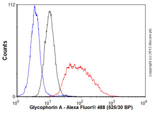
Overlay histogram showing K562 cells stained with unpurified ab129024 (red line). The cells were fixed with 80% methanol (5 min) and then permeabilized with 0.1% PBS-Tween for 20 min. The cells were then incubated in 1x PBS / 10% normal goat serum / 0.3M glycine to block non-specific protein-protein interactions followed by the antibody (ab129024, 1/100 dilution) for 30 min at 22°C. The secondary antibody used was Alexa Fluor® 488 goat anti-rabbit IgG (H&L) (ab150077) at 1/2000 dilution for 30 min at 22°C. Isotype control antibody (black line) was rabbit IgG (monoclonal) (1μg/1x106 cells) used under the same conditions. Unlabelled sample (blue line) was also used as a control. Acquisition of >5,000 events were collected using a 20mW Argon ion laser (488nm) and 525/30 bandpass filter.
![Anti-Glycophorin A antibody [EPR8200] (ab129024) at 1/1000 dilution (unpurified) + Fetal liver lysate at 10 µg](http://www.bioprodhub.com/system/product_images/ab_products/2/sub_2/25825_Glycophorin-A-Primary-antibodies-ab129024-1.jpg)
Anti-Glycophorin A antibody [EPR8200] (ab129024) at 1/1000 dilution (unpurified) + Fetal liver lysate at 10 µg
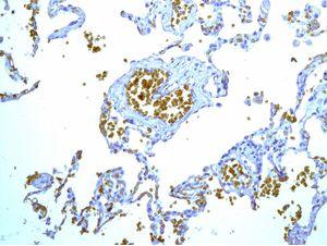
ab129024, at 1/100 dilution staining Glycophorin A in formalin fixed paraffin embedded Human lung tissue by immunohistochemistry.
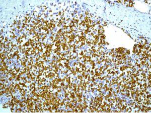
ab129024, unpurified, at 1/100 dilution staining Glycophorin A in formalin fixed paraffin embedded Human spleen tissue by immunohistochemistry.
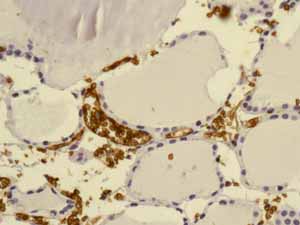
ab129024, unpurified, showing positive staining in Thyroid gland erythrocytes tissue.
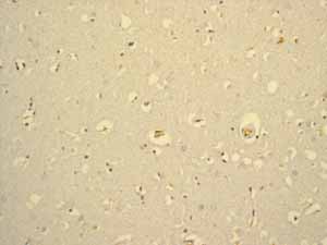
ab129024, unpurified, showing negative staining in Normal brain tissue.
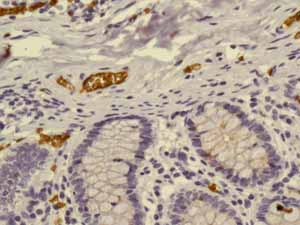
ab129024, unpurified, showing positive staining in Normal colon erythrocytes tissue.
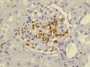
ab129024, unpurified, showing positive staining in Normal kidney erythrocytes tissue.
ab129024, unpurified, showing positive staining in Normal placenta erythrocytes tissue.
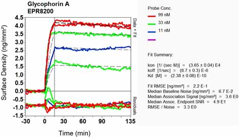
Equilibrium disassociation constant (KD)Learn more about KD Click here to learn more about KD