![All lanes : Anti-GLO1 antibody [Glo1a] (ab171121) at 1/1000 dilutionLane 1 : HeLa cell lysateLane 2 : HepG2 cell lysateLane 3 : 293T cell lysateLane 4 : A431 cell lysateLane 5 : A549 cell lysateLane 6 : MCF7 cell lysateLane 7 : U2OS cell lysateLane 8 : K562 cell lysateLane 9 : monkey COS7 cell lysateLane 10 : mouse MEF cell lysateLane 11 : mouse C2C12 cell lysateLane 12 : rat NRK cell lysateLysates/proteins at 25 µg per lane.Secondarygoat anti-mouse-HRP at 1/20000 dilution](http://www.bioprodhub.com/system/product_images/ab_products/2/sub_2/24848_ab171121-171275-ab171121WB2.jpg)
All lanes : Anti-GLO1 antibody [Glo1a] (ab171121) at 1/1000 dilutionLane 1 : HeLa cell lysateLane 2 : HepG2 cell lysateLane 3 : 293T cell lysateLane 4 : A431 cell lysateLane 5 : A549 cell lysateLane 6 : MCF7 cell lysateLane 7 : U2OS cell lysateLane 8 : K562 cell lysateLane 9 : monkey COS7 cell lysateLane 10 : mouse MEF cell lysateLane 11 : mouse C2C12 cell lysateLane 12 : rat NRK cell lysateLysates/proteins at 25 µg per lane.Secondarygoat anti-mouse-HRP at 1/20000 dilution
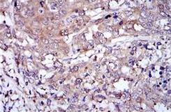
Immunohistochemical analysis of deparaffinized Human stomach cancer tissue, labeling GLO1 with ab171121 at 1/800 dilution. Colorimetric detection was performed using metal enhanced DAB and tissues counterstained with hematoxylin.

Immunohistochemical analysis of deparaffinized Human prostate cancer tissue, labeling GLO1 with ab171121 at 1/800 dilution. Colorimetric detection was performed using metal enhanced DAB and tissues counterstained with hematoxylin.