Anti-GFAP antibody - Astrocyte Marker
| Name | Anti-GFAP antibody - Astrocyte Marker |
|---|---|
| Supplier | Abcam |
| Catalog | ab48050 |
| Prices | $398.00 |
| Sizes | 100 µg |
| Host | Rabbit |
| Clonality | Polyclonal |
| Isotype | IgG |
| Applications | ELISA IHC-F IHC-P ICC/IF ICC/IF WB |
| Species Reactivities | Mouse, Rat, Sheep, Goat, Human, Monkey |
| Antigen | Synthetic peptide conjugated to KLH derived from within residues 400 to the C-terminus of Human GFAP |
| Blocking Peptide | Human GFAP peptide |
| Description | Rabbit Polyclonal |
| Gene | GFAP |
| Conjugate | Unconjugated |
| Supplier Page | Shop |
Product images
![Standard Curve for GFAP; dilution range 1 pg/ml to 1 ug/ml using Capture Antibody Mouse monoclonal [2A5] to GFAP - Astrocyte Marker (ab4648) at 0.2 ug/ml and Detector Antibody Rabbit polyclonal to GFAP - Astrocyte Marker (ab48050) at 0.5 ug/ml.](http://www.bioprodhub.com/system/product_images/ab_products/2/sub_2/24109_GFAP-Primary-antibodies-ab48050-8.jpg) Standard Curve for GFAP; dilution range 1 pg/ml to 1 ug/ml using Capture Antibody Mouse monoclonal [2A5] to GFAP - Astrocyte Marker (ab4648) at 0.2 ug/ml and Detector Antibody Rabbit polyclonal to GFAP - Astrocyte Marker (ab48050) at 0.5 ug/ml.
Standard Curve for GFAP; dilution range 1 pg/ml to 1 ug/ml using Capture Antibody Mouse monoclonal [2A5] to GFAP - Astrocyte Marker (ab4648) at 0.2 ug/ml and Detector Antibody Rabbit polyclonal to GFAP - Astrocyte Marker (ab48050) at 0.5 ug/ml.
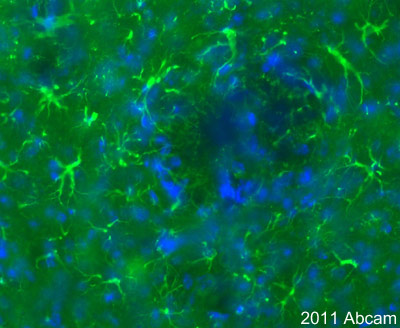 Immunoflourescent staining of mouse brain using anti-GFAP (ab 48050). Mouse tissues were fixed (animals perfused) with 4% PFA and later postfixed overnight in the same fixative. They were cryoprotected in 30% sucrose and cut with microtome (25 µm thickness). Immunofuorescent staining performed on mouse brain sections blocked with 10% serum over 1 hour using the anti-GFAP primary antibody (ab 48050) at 1/200 for 24 hours at 4°C. Incubation with anti-rabbit Alexa 488 at 1/1000 followed. The antibody stains astrocytes and nucleus stained with Hoechst at high power magnification.See Abreview
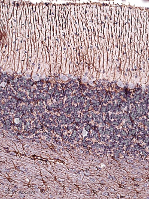 Immunohistochemical staining of rat cerebellum using anti-GFAP (ab 48050). Tissues were fixed with formaldehyde and antigen retrieved using heat mediated citric acid buffer. Immunohistochemical analysis was performed on rat cerebellum sections (blocked with 1% BSA for 10 minutes) using the anti-GFAP primary antibody (ab48050) at 1/2000 for 2 hours at 21°C. This was followed by incubation with anti-rabbit biotin conjugated secondary anbibody at 1/200. Astrocytes appear to be specifically immunolabelled.See Abreview
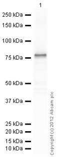
 All lanes : Anti-GFAP antibody - Astrocyte Marker (ab48050) at 1 µg/mlLane 1 : Brain (Human) Tissue Lysate - adult normal tissue (ab29466)Lane 2 : Brain (Rat) Rat Tissue LysateLysates/proteins at 10 µg per lane.SecondaryIRDye 680 Conjugated Goat Anti-Rabbit IgG (H+L) at 1/10000 dilutionPerformed under reducing conditions.
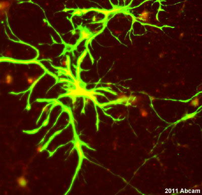 Fluorescent immunocytochemistry staining of cultured rat astrocytes using anti-GFAP (ab 48050). Cultured rat astrocytes were fixed with 4% PFA and permeabilized with 0.1% Triton X in 0.1% PBS. Fluorescent immunocytochemistry staining performed on cultured rat astrocytes blocked with 10% serum over 1 hour at 24ºC using anti-GFAP primary antibody (ab 48050) at 1/200 for 4 hours at 24°C. Incubation with donkey anti-rabbit Alexa 488 secondary at 1/1000 followed. Cells co-stained with BMP6 in red.See Abreview
 Anti-GFAP antibody - Astrocyte Marker (ab48050) at 1 µg/ml + Human GFAP full length protein (ab114149) at 0.1 µgSecondaryGoat Anti-Rabbit IgG H&L (HRP) preadsorbed (ab97080) at 1/5000 dilutiondeveloped using the ECL techniquePerformed under reducing conditions.Exposure time : 10 seconds
Product References
Potentiation of cytotoxic chemotherapy by growth hormone-releasing hormone - Potentiation of cytotoxic chemotherapy by growth hormone-releasing hormone
Jaszberenyi M, Rick FG, Popovics P, Block NL, Zarandi M, Cai RZ, Vidaurre I, Szalontay L, Jayakumar AR, Schally AV. Proc Natl Acad Sci U S A. 2014 Jan 14;111(2):781-6.
Localisation of citrullinated proteins in normal appearing white matter and - Localisation of citrullinated proteins in normal appearing white matter and
Bradford CM, Ramos I, Cross AK, Haddock G, McQuaid S, Nicholas AP, Woodroofe MN. J Neuroimmunol. 2014 Aug 15;273(1-2):85-95.
Circulating neprilysin clears brain amyloid. - Circulating neprilysin clears brain amyloid.
Liu Y, Studzinski C, Beckett T, Murphy MP, Klein RL, Hersh LB. Mol Cell Neurosci. 2010 Oct;45(2):101-7.
Time-dependent changes in ghrelin-immunoreactivity in dissociated neuronal - Time-dependent changes in ghrelin-immunoreactivity in dissociated neuronal
Stoyanova II, Wiertz RW, Rutten WL. Regul Pept. 2009 Nov 27;158(1-3):86-90.