Anti-GFAP antibody [EP672Y]
| Name | Anti-GFAP antibody [EP672Y] |
|---|---|
| Supplier | Abcam |
| Catalog | ab33922 |
| Prices | $398.00 |
| Sizes | 100 µl |
| Host | Rabbit |
| Clonality | Monoclonal |
| Isotype | IgG |
| Clone | EP672Y |
| Applications | ICC/IF ICC/IF IHC-F WB IHC-P ICC/IF |
| Species Reactivities | Rat, Human |
| Antigen | Synthetic peptide (the amino acid sequence is considered to be commercially sensitive) (C terminal) |
| Description | Rabbit Monoclonal |
| Gene | GFAP |
| Conjugate | Unconjugated |
| Supplier Page | Shop |
Product images
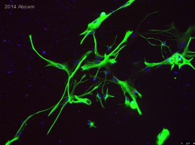 ICC/IF image of ab33922 stained Rat primary mixed astrocytes culture. The cells were 100% Paraformaldehyde fixed and then incubated in 10% Serum / 0.1M PBS with 10% Donkey serum for 4h. The secondary antibody was Alexa Fluor® 488 goat anti-rabbit IgG (H+L) used at a 1/1000 dilution.
![Anti-GFAP antibody [EP672Y] (ab33922) at 1/2000 dilution + Rat brain lysate](http://www.bioprodhub.com/system/product_images/ab_products/2/sub_2/24084_33922.jpg) Anti-GFAP antibody [EP672Y] (ab33922) at 1/2000 dilution + Rat brain lysate
Anti-GFAP antibody [EP672Y] (ab33922) at 1/2000 dilution + Rat brain lysate
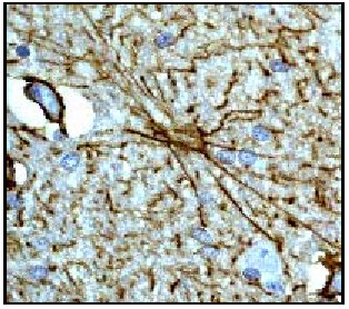 Immjunohistochemical analysis of paraffin-embedded human brain using ab33922 at 1/250.Immjunohistochemical analysis of paraffin-embedded human brain using ab33922 at 1/250.
 ab33922 staining GFAP in a xenograft section, human neural stem cells in fetal mouse, by Immunohistochemistry (Frozen sections).Tissue was fixed in paraformaldehyde and permeabilized with 0.2% Triton X. Samples were then blocked using 3% normal donkey serum for 1.5 hours at 20°C, then incubated with ab33922 at a 1/1000 dilution for 1 hour at 20°C. The secondary used was a donkey anti-rabbit polyclonal conjugated to Alexa Fluor 555, used at a 1/500 dilution.ab33922 only recognizes human astrocytes and none of the murine tissue was stained. The staining of the human GFAP fibers was always very strong, clear and reliable. Very good antibody for xenograft experiments in neuroscience!See Abreview
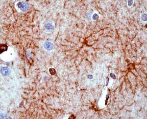 ab33922 showing positive staining in Normal brain tissue.
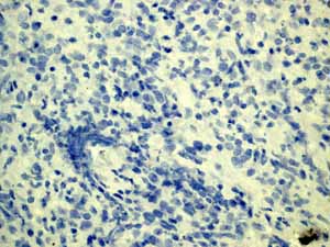
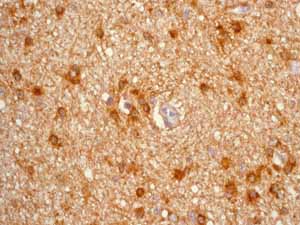 ab33922 showing positive staining in Astrocytoma tissue.
 ab33922 showing negative staining in Meningioma tissue.
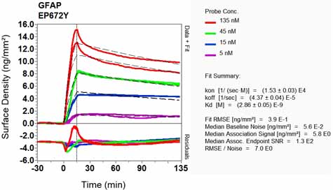 Equilibrium disassociation constant (KD)Learn more about KD Click here to learn more about KD
Product References
Hepatitis B virus infection and immunopathogenesis in a humanized mouse model: - Hepatitis B virus infection and immunopathogenesis in a humanized mouse model:
Bility MT, Cheng L, Zhang Z, Luan Y, Li F, Chi L, Zhang L, Tu Z, Gao Y, Fu Y, Niu J, Wang F, Su L. PLoS Pathog. 2014 Mar 20;10(3):e1004032.
Neurite outgrowth and differentiation of rat cortex progenitor cells are - Neurite outgrowth and differentiation of rat cortex progenitor cells are
Jeerage KM, Oreskovic TL, Hume SL. Neurotoxicology. 2012 Oct;33(5):1170-9.