![Overlay histogram showing SHSY-5Y cells stained with ab106509 (red line). The cells were fixed with 4% paraformaldehyde (10 min) and then permeabilized with 0.1% PBS-Tween for 20 min. The cells were then incubated in 1x PBS / 10% normal goat serum / 0.3M glycine to block non-specific protein-protein interactions followed by the antibody (ab106509, 1/1000 dilution) for 30 min at 22°C. The secondary antibody used was Alexa Fluor® 488 goat anti-mouse IgG (H&L) (ab150113) at 1/2000 dilution for 30 min at 22°C. Isotype control antibody (black line) was mouse IgG1 [ICIGG1] (ab91353, 1μg/1x106 cells) used under the same conditions. Unlabelled sample (blue line) was also used as a control. Acquisition of >5,000 events were collected using a 20mW Argon ion laser (488nm) and 525/30 bandpass filter. This antibody gave a positive signal in SHSY-5Y cells fixed with 80% methanol (5 min)/permeabilized with 0.1% PBS-Tween for 20 min used under the same conditions.](http://www.bioprodhub.com/system/product_images/ab_products/2/sub_2/24078_ab106509-4-ab106509FC.jpg)
Overlay histogram showing SHSY-5Y cells stained with ab106509 (red line). The cells were fixed with 4% paraformaldehyde (10 min) and then permeabilized with 0.1% PBS-Tween for 20 min. The cells were then incubated in 1x PBS / 10% normal goat serum / 0.3M glycine to block non-specific protein-protein interactions followed by the antibody (ab106509, 1/1000 dilution) for 30 min at 22°C. The secondary antibody used was Alexa Fluor® 488 goat anti-mouse IgG (H&L) (ab150113) at 1/2000 dilution for 30 min at 22°C. Isotype control antibody (black line) was mouse IgG1 [ICIGG1] (ab91353, 1μg/1x106 cells) used under the same conditions. Unlabelled sample (blue line) was also used as a control. Acquisition of >5,000 events were collected using a 20mW Argon ion laser (488nm) and 525/30 bandpass filter. This antibody gave a positive signal in SHSY-5Y cells fixed with 80% methanol (5 min)/permeabilized with 0.1% PBS-Tween for 20 min used under the same conditions.
![All lanes : Anti-GFAP antibody [6A6] (ab106509) at 1/200 dilutionLane 1 : A431 cell lysate Lane 2 : SK-N-SH cell lysate Lane 3 : PC12 cell lysate 20ugLysates/proteins at 20 µg per lane.](http://www.bioprodhub.com/system/product_images/ab_products/2/sub_2/24079_GFAP-Primary-antibodies-ab106509-2.jpg)
All lanes : Anti-GFAP antibody [6A6] (ab106509) at 1/200 dilutionLane 1 : A431 cell lysate Lane 2 : SK-N-SH cell lysate Lane 3 : PC12 cell lysate 20ugLysates/proteins at 20 µg per lane.
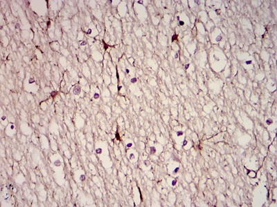
Immunohistochemical analysis of paraffin-embedded human brain tissues using ab106509 at a dilution of 1/200 with DAB staining.
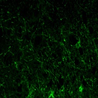
Immunofluorescence analysis of paraffin-embedded lobe of brain tissues using ab106509 at a dilution of 1/200 (green).