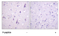
ab62479, at 1/50 dilution, staining GFAP in paraffin embedded human brain tissue by Immunohistochemsitry in the absence (left image) or presence (right image) of the immunising peptide.

ab62479 staining GFAP (phospho S38) in mouse brain tissue section by Immunohistochemistry (Frozen sections). Tissue samples were fixed with paraformaldehyde and blocking with a dilution buffer containing 2% Block Ace, 0.05% Triton X-100 and PBS was performed at 250C for 1 hour. The sample was incubated with primary antibody (1/100) at 40C for 12 hours. A Cyt3 ® -conjugated donkey polyclonal to rabbit IgG was used as secondary antibody at 1/500 dilution.See Abreview