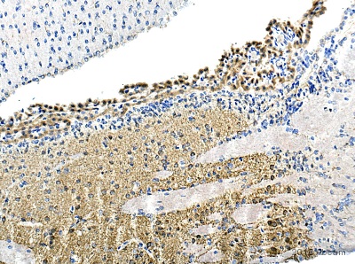
IHC-P image of Gephyrin staining on rat Caudate putamen sections using ab136343 (1:100). The sections were deparaffinized and subjected to heat mediated antigen retrieval uisng citric acid. The sections were then blocked using 1% BSA for 10 mins at 21°C. ab136343 was diluted 1:100 and incubated with the section for 16 hours at 21°C. The secondary antibody used was ab6876 - Goat polyclonal to Chicken IgY H&L conjugated to Biotin (1:200)
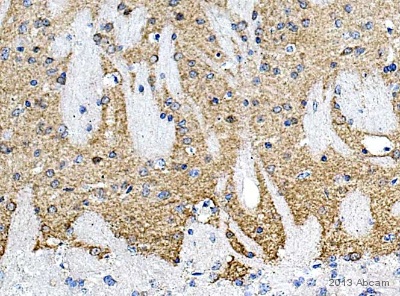
IHC-P image of Gephyrin staining on mouse Caudate putamen sections using ab136343 (1:100). The sections were deparaffinized and subjected to heat mediated antigen retrieval uisng citric acid. The sections were then blocked using 1% BSA for 10 mins at 21°C. ab136343 was diluted 1:100 and incubated with the section for 16 hours at 21°C. The secondary antibody used was ab6876 - Goat polyclonal to Chicken IgY H&L conjugated to Biotin (1:200)
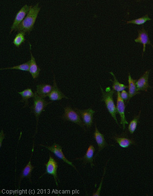
ICC/IF image of ab136343 stained MEF1 cells. The cells were 4% formaldehyde fixed (10 min) and then incubated in 1%BSA / 10% normal goat serum / 0.3M glycine in 0.1% PBS-Tween for 1h to permeabilise the cells and block non-specific protein-protein interactions. The cells were then incubated with the antibody (ab136434, 5µg/ml) overnight at +4°C. The secondary antibody (green) was ab96947, DyLight® 488 goat anti-chicken IgY (H+L) used at a 1/250 dilution for 1h. Alexa Fluor® 594 WGA was used to label plasma membranes (red) at a 1/200 dilution for 1h. DAPI was used to stain the cell nuclei (blue) at a concentration of 1.43µM.
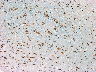
IHC image of Gephyrin staining in Rat brain formalin fixed paraffin embedded tissue section, performed on a Leica BondTM system using the standard protocol B. The section was pre-treated using heat mediated antigen retrieval with sodium citrate buffer (pH6, epitope retrieval solution 1) for 20 mins. The section was then incubated with ab136343, 5µg/ml, for 15 mins at room temperature. A Goat anti-Chicken biotinylated secondary antibody was used to detect the primary, and visualized using an HRP conjugated ABC system. DAB was used as the chromogen. The section was then counterstained with haematoxylin and mounted with DPX. For other IHC staining systems (automated and non-automated) customers should optimize variable parameters such as antigen retrieval conditions, primary antibody concentration and antibody incubation times.