![All lanes : Anti-GCAT antibody [EPR13450] - N-terminal (ab181094) at 1/5000 dilutionLane 1 : BxPC-3 cell lysateLane 2 : HepG2 cell lysateLane 3 : U87-MG cell lysateLysates/proteins at 20 µg per lane.SecondaryGoat Anti-Rabbit IgG, (H+L), Peroxidase conjugated at 1/1000 dilution](http://www.bioprodhub.com/system/product_images/ab_products/2/sub_2/23386_ab181094-214730-ab181094WB1.jpg)
All lanes : Anti-GCAT antibody [EPR13450] - N-terminal (ab181094) at 1/5000 dilutionLane 1 : BxPC-3 cell lysateLane 2 : HepG2 cell lysateLane 3 : U87-MG cell lysateLysates/proteins at 20 µg per lane.SecondaryGoat Anti-Rabbit IgG, (H+L), Peroxidase conjugated at 1/1000 dilution
![Anti-GCAT antibody [EPR13450] - N-terminal (ab181094) at 1/5000 dilution + Human fetal liver lysateSecondaryGoat Anti-Rabbit IgG H&L (HRP) (ab136636) at 1/500 dilution](http://www.bioprodhub.com/system/product_images/ab_products/2/sub_2/23387_ab181094-214731-ab181094WB2.jpg)
Anti-GCAT antibody [EPR13450] - N-terminal (ab181094) at 1/5000 dilution + Human fetal liver lysateSecondaryGoat Anti-Rabbit IgG H&L (HRP) (ab136636) at 1/500 dilution
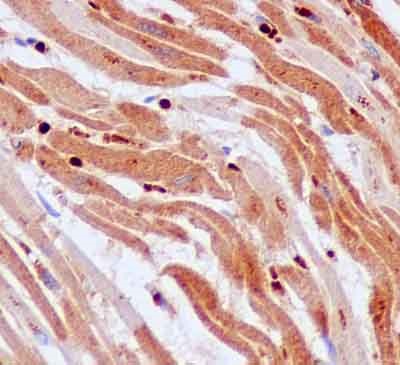
Immunohistochemical analysis of Human heart tissue, staining GCAT with ab181094 at 1/100 dilution. Detected using HRP Polymer for Rabbit IgG and counter-stained using hematoxylin.
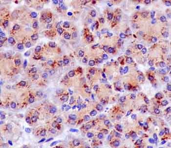
Immunohistochemical analysis of Human pancreas tissue, staining GCAT with ab181094 at 1/100 dilution. Detected using HRP Polymer for Rabbit IgG and counter-stained using hematoxylin.
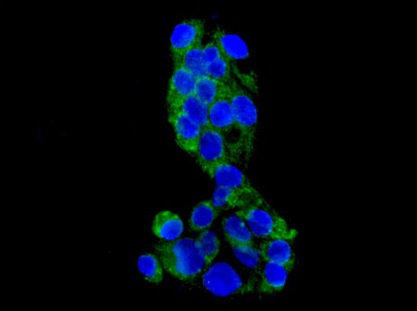
Immunofluorescence analysis of HepG2 cells, staining GCAT (green) with ab181094 at 1/100 dilution. An Alexa Fluor®488-conjugated goat anti-rabbit IgG was used as the secondary antibody at 1/200 dilution. Nuclei were counterstained with DAPI (blue).
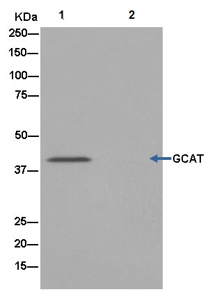
Western blot analysis on immunoprecipitation pellet from HepG2 cell lysate (lane 1) or negative control (lane 2), labeling GCAT immunoprecipitated using ab181094 at 1/40 dilution and HRP-conjugated anti-rabbit IgG preferentially detecting the non-reduced form of rabbit IgG.