![Anti-GATA1 antibody [4F5] (ab98953) at 1/500 dilution + K562 cell lysate](http://www.bioprodhub.com/system/product_images/ab_products/2/sub_2/23005_GATA1-Primary-antibodies-ab98953-1.jpg)
Anti-GATA1 antibody [4F5] (ab98953) at 1/500 dilution + K562 cell lysate

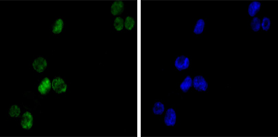
Left: immunofluorescence analysis of K562 cells using ab98953 diluted 1/200 (green). Right: DRAQ5 fluorescent DNA dye (blue).

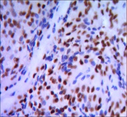
Immunohistochemical analysis of paraffin-embedded pancreatic cancer tissue, using ab98953 at 1/200 dilution with DAB staining.

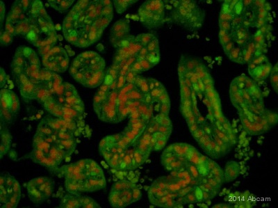
ab98953 staining GATA1 (green) in Baboon placenta tissue sections by Immunohistochemistry (IHC-P - paraformaldehyde-fixed, paraffin-embedded sections). Tissue was fixed with formaldehyde and blocked with 5% serum for 30 minutes at 22°C; antigen retrieval was by heat mediation in 10mM sodium citrate buffer, pH 6.0. Samples were incubated with primary antibody (1/200 in PBS + 5% normal goat serum) for 16 hours at 4°C. A Biotin-conjugated goat anti-mouse IgG (H+L) polyclonal (7µg/ml) was used as the secondary antibody. Red = propidium iodide stained nuclei.See Abreview
![Anti-GATA1 antibody [4F5] (ab98953) at 1/500 dilution + K562 cell lysate](http://www.bioprodhub.com/system/product_images/ab_products/2/sub_2/23005_GATA1-Primary-antibodies-ab98953-1.jpg)


