![All lanes : Anti-GART antibody [EPR11622] (ab169550) at 1/1000 dilutionLane 1 : HeLa cell lysateLane 2 : HepG2 cell lysateLane 3 : K562 cell lysateLane 4 : A431 cell lysateLysates/proteins at 10 µg per lane.SecondaryHRP labeled goat anti-rabbit at 1/2000 dilution](http://www.bioprodhub.com/system/product_images/ab_products/2/sub_2/22896_GART-Primary-antibodies-ab169550-1.jpg)
All lanes : Anti-GART antibody [EPR11622] (ab169550) at 1/1000 dilutionLane 1 : HeLa cell lysateLane 2 : HepG2 cell lysateLane 3 : K562 cell lysateLane 4 : A431 cell lysateLysates/proteins at 10 µg per lane.SecondaryHRP labeled goat anti-rabbit at 1/2000 dilution
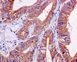
Immunohistochemical analysis of paraffin-embedded Human colonic adenocarcinoma tissue labeling GART with ab169550 at 1/50 dilution.
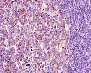
Immunohistochemical analysis of paraffin-embedded Human tonsil tissue labeling GART with ab169550 at 1/50 dilution.
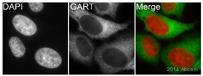
ab169550 staining GART in human HeLa cells by ICC/IF (Immunocytochemistry/immunofluorescence). Cells were fixed with paraformaldehyde and permeabilized with 0.5% Triton X-100 in PBS. Samples were incubated with primary antibody (1/500 in PBS) for 1 hour at 22°C. An Alexa Fluor® 488-conjugated goat anti-rabbit IgG polyclonal (1/200) was used as the secondary antibody. Counterstained with DAPI.See Abreview
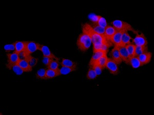
Immunofluorescent analysis of HepG2 cells labeling GART with ab169550 at 1/100 dilution.
![Anti-GART antibody [EPR11622] (ab169550) at 1/1000 dilution + immunoprecipitation pellet from HeLa cell lysateSecondaryHRP-conjugated anti-rabbit IgG preferentially detecting the non-reduced form of rabbit IgG](http://www.bioprodhub.com/system/product_images/ab_products/2/sub_2/22901_GART-Primary-antibodies-ab169550-6.jpg)
Anti-GART antibody [EPR11622] (ab169550) at 1/1000 dilution + immunoprecipitation pellet from HeLa cell lysateSecondaryHRP-conjugated anti-rabbit IgG preferentially detecting the non-reduced form of rabbit IgG