![All lanes : Anti-gamma Tubulin antibody [4D11] - C-terminal (ab173831) at 1/1000 dilutionLane 1 : HeLa cell lysateLane 2 : HepG2 cell lysateLane 3 : U2OS cell lysateLane 4 : K562 cell lysateLane 5 : mouse 3T3L1 cell lysateLane 6 : mouse spleen lysateLane 7 : monkey COS7 cell lysateLysates/proteins at 50 µg per lane.Secondarygoat anti-mouse-HRP at 1/20000 dilutiondeveloped using the ECL technique](http://www.bioprodhub.com/system/product_images/ab_products/2/sub_2/22595_ab173831-197561-ab173831WB1.jpg)
All lanes : Anti-gamma Tubulin antibody [4D11] - C-terminal (ab173831) at 1/1000 dilutionLane 1 : HeLa cell lysateLane 2 : HepG2 cell lysateLane 3 : U2OS cell lysateLane 4 : K562 cell lysateLane 5 : mouse 3T3L1 cell lysateLane 6 : mouse spleen lysateLane 7 : monkey COS7 cell lysateLysates/proteins at 50 µg per lane.Secondarygoat anti-mouse-HRP at 1/20000 dilutiondeveloped using the ECL technique
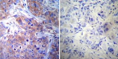
Immunohistochemical analysis of Human breast carcinoma tissue, labeling gamma Tubulin with ab173831 at a 1/20 dilution (left panel) or without primary antibody (right panel). To expose target proteins, heat induced antigen retrieval was performed using 10mM sodium citrate (pH6.0) buffer, microwaved for 8-15 minutes. Detection was performed using a biotin-conjugated secondary antibody and SA-HRP, followed by colorimetric detection using DAB. Tissues were counterstained with hematoxylin and prepped for mounting.
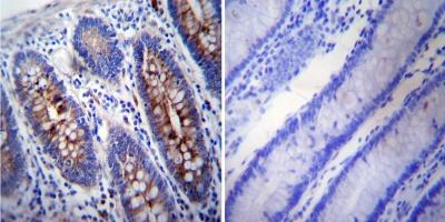
Immunohistochemical analysis of Human colon tissue, labeling gamma Tubulin with ab173831 at a 1/200 dilution (left panel) or without primary antibody (right panel). To expose target proteins, heat induced antigen retrieval was performed using 10mM sodium citrate (pH6.0) buffer, microwaved for 8-15 minutes. Detection was performed using a biotin-conjugated secondary antibody and SA-HRP, followed by colorimetric detection using DAB. Tissues were counterstained with hematoxylin and prepped for mounting.
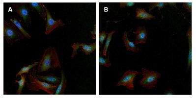
Immunofluorescence analysis of formalin-fixed, permeabilized A) HeLa cells and B) U2OS, labeling gamma Tubulin (green) using ab173831 at a 1/50 dilution. Cells were washed with PBS and incubated with a DyLight-488 conjugated secondary antibody. F-Actin (red) was stained with DyLight 547 Phalloidin and nuclei (blue) were stained with Hoechst 33342 dye.