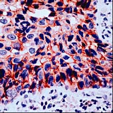
Human breast carcinoma stained with anti gamma catenin antibody ab15153 (1/100 for 10 min at RT). Gamma catenin staining is seen on the cell membrane and in the cytoplasm.

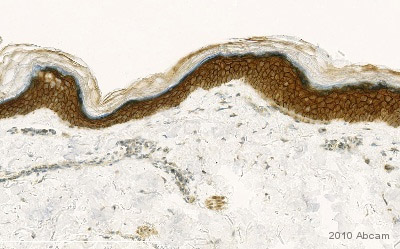
ab15153 staining gamma Catenin in normal human skin tissue sections by IHC-P (formaldehyde-fixed paraffin-embedded sections). Tissue samples were fixed with formaldehyde and blocked with 10% serum for 1 hour at 2°C; antigen retrival was by heat mediation in EDTA (pH9). The sample was incubated with primary antibody (1/100 in TBS + 1% BSA) at 21°C for 1 hour. An undiluted HRP-conjugated Goat polyclonal to mouse IgG was used as secondary antibody.See Abreview

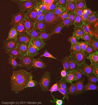
ICC/IF image of ab15153 stained MCF7 cells. The cells were 100% methanol fixed (5 min) and then incubated in 1%BSA / 10% normal goat serum / 0.3M glycine in 0.1% PBS-Tween for 1h to permeabilise the cells and block non-specific protein-protein interactions. The cells were then incubated with the antibody (ab15153, 1µg/ml) overnight at +4°C. The secondary antibody (green) was Alexa Fluor® 488 goat anti-rabbit IgG (H+L) used at a 1/1000 dilution for 1h. Alexa Fluor® 594 WGA was used to label plasma membranes (red) at a 1/200 dilution for 1h. DAPI was used to stain the cell nuclei (blue) at a concentration of 1.43µM.


