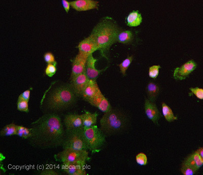
ICC/IF image of ab76245 stained Panc-1 cells. The cells were 4% formaldehyde fixed (10 min) and then incubated in 1%BSA / 10% normal goat serum / 0.3M glycine in 0.1% PBS-Tween for 1h to permeabilise the cells and block non-specific protein-protein interactions. The cells were then incubated with the antibody ab76245 at 10µg/ml overnight at +4°C. The secondary antibody (pseudo-colored green) was Alexa Fluor® 488 goat anti- rabbit (ab150081) IgG (H+L) preadsorbed, used at a 1/1000 dilution for 1h. Alexa Fluor® 594 WGA was used to label plasma membranes (pseudo-colored red) at a 1/200 dilution for 1h at room temperature. DAPI was used to stain the cell nuclei (pseudo-colored blue) at a concentration of 1.43µM for 1hour at room temperature.
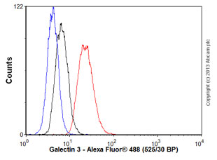
Overlay histogram showing THP1 cells stained with ab76245 (red line). The cells were fixed with 80% methanol (5 min) and then permeabilized with 0.1% PBS-Tween for 20 min. The cells were then incubated in 1x PBS / 10% human serum / 0.3M glycine to block non-specific protein-protein interactions followed by the antibody (ab76245, 1/1000 dilution) for 30 min at 22°C. The secondary antibody used was Alexa Fluor® 488 goat anti-rabbit IgG (H&L) (ab150077) at 1/2000 dilution for 30 min at 22°C. Isotype control antibody (black line) was rabbit IgG (monoclonal) (0.1μg/1x106 cells) used under the same conditions. Unlabelled sample (blue line) was also used as a control. Acquisition of >5,000 events were collected using a 20mW Argon ion laser (488nm) and 525/30 bandpass filter.
![All lanes : Anti-Galectin 3 antibody [EP2775Y] (ab76245) at 1/10000 dilutionLane 1 : A375 cell lysateLane 2 : HeLa cell lysateLane 3 : SW480 cell lysateLane 4 : A431 cell lysateLysates/proteins at 10 µg per lane.SecondaryHRP labelled goat anti rabbit at 1/2000 dilution](http://www.bioprodhub.com/system/product_images/ab_products/2/sub_2/22247_ab76245_WB_1.jpg)
All lanes : Anti-Galectin 3 antibody [EP2775Y] (ab76245) at 1/10000 dilutionLane 1 : A375 cell lysateLane 2 : HeLa cell lysateLane 3 : SW480 cell lysateLane 4 : A431 cell lysateLysates/proteins at 10 µg per lane.SecondaryHRP labelled goat anti rabbit at 1/2000 dilution
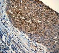
Immunohistochemical analysis of paraffin-embedded human lung squamous carcinoma using anti-Galectin-3 RabMAb (ab76245).