Anti-Galectin 3 antibody [A3A12]
| Name | Anti-Galectin 3 antibody [A3A12] |
|---|---|
| Supplier | Abcam |
| Catalog | ab2785 |
| Prices | $403.00 |
| Sizes | 100 µg |
| Host | Mouse |
| Clonality | Monoclonal |
| Isotype | IgG1 |
| Clone | A3A12 |
| Applications | ELISA IHC-F WB ICC/IF ICC/IF B/N IHC-P ICC/IF |
| Species Reactivities | Mouse, Rat, Rabbit, Human |
| Antigen | Recombinant full length protein corresponding to Human Galectin 3 |
| Description | Mouse Monoclonal |
| Gene | LGALS3 |
| Conjugate | Unconjugated |
| Supplier Page | Shop |
Product images
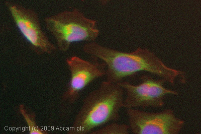
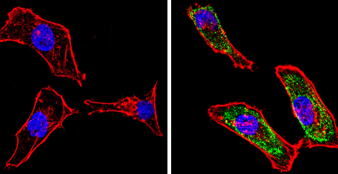 ab2785 labelling Galectin 3 (green) in the cytoplasm and nucleus of HeLa cells by Immunocytochemistry/Immunofluorescence. Formalin-fixed cells were permeabilized with 0.1% Triton X-100 in TBS for 5-10 minutes and blocked with 3% BSA-PBS for 30 minutes at room temperature. Cells were incubated with the primary antibody (1:100 in 3% BSA-PBS) overnight at 4 ºC. A DyLight-conjugated anti-mouse was used as the secondary antibody. Red (phalloidin) - F-actin, Blue - nuclei. Images were taken at a magnification of 60x.
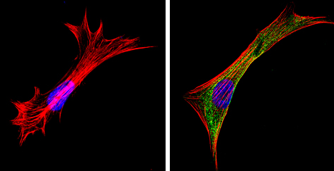 ab2785 labelling Galectin 3 (green) in the cytoplasm and nucleus of NIH-3T3 cells by Immunocytochemistry/Immunofluorescence. Formalin-fixed cells were permeabilized with 0.1% Triton X-100 in TBS for 5-10 minutes and blocked with 3% BSA-PBS for 30 minutes at room temperature. Cells were incubated with the primary antibody (1:100 in 3% BSA-PBS) overnight at 4 ºC. A DyLight-conjugated anti-mouse was used as the secondary antibody. Red (phalloidin) - F-actin, Blue - nuclei. Images were taken at a magnification of 60x.
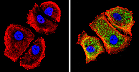 ab2785 labelling Galectin 3 (green) in the cytoplasm and nucleus of MCF-7 cells by Immunocytochemistry/Immunofluorescence. Formalin-fixed cells were permeabilized with 0.1% Triton X-100 in TBS for 5-10 minutes and blocked with 3% BSA-PBS for 30 minutes at room temperature. Cells were incubated with the primary antibody (1:100 in 3% BSA-PBS) overnight at 4 ºC. A DyLight-conjugated anti-mouse was used as the secondary antibody. Red (phalloidin) - F-actin, Blue - nuclei. Images were taken at a magnification of 60x.
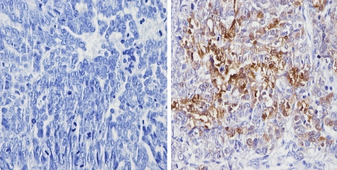 ab2785 labelling Galectin 3 in the nucleus and cytoplasm of Human ovarian carcinoma tissue (right) compared with a negative control (left) by Immunohistochemistry (formalin/PFA-fixed paraffin-embedded sections). To expose target proteins, antigen retrieval method was performed using 10mM sodium citrate (pH 6.0) microwaved for 8-15 min. Following antigen retrieval, tissues were blocked in 3% H2O2-methanol for 15 min at room temperature. Tissue sections were incubated with the primary antibody (1:100 in 3% BSA-PBS) overnight at 4°C. A HRP-conjugated anti-mouse IgG was used as the secondary antibody, followed by colorimetric detection using a DAB kit. Tissues were counterstained with hematoxylin and dehydrated with ethanol and xylene to prep for mounting.
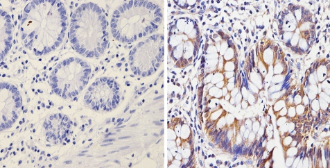 ab2785 labelling Galectin 3 in the nucleus and cytoplasm of Human colon tissue (right) compared with a negative control (left) by Immunohistochemistry (formalin/PFA-fixed paraffin-embedded sections). To expose target proteins, antigen retrieval method was performed using 10mM sodium citrate (pH 6.0) microwaved for 8-15 min. Following antigen retrieval, tissues were blocked in 3% H2O2-methanol for 15 min at room temperature. Tissue sections were incubated with the primary antibody (1:100 in 3% BSA-PBS) overnight at 4°C. A HRP-conjugated anti-mouse IgG was used as the secondary antibody, followed by colorimetric detection using a DAB kit. Tissues were counterstained with hematoxylin and dehydrated with ethanol and xylene to prep for mounting.
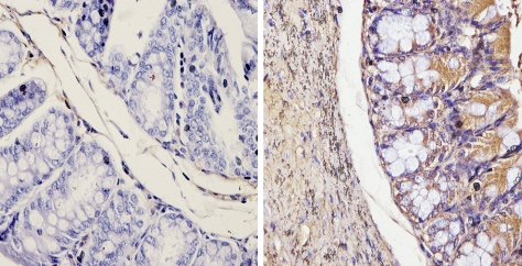 ab2785 labelling Galectin 3 in the nucleus and cytoplasm of Mouse colon tissue (right) compared with a negative control (left) by Immunohistochemistry (formalin/PFA-fixed paraffin-embedded sections). To expose target proteins, antigen retrieval method was performed using 10mM sodium citrate (pH 6.0) microwaved for 8-15 min. Following antigen retrieval, tissues were blocked in 3% H2O2-methanol for 15 min at room temperature. Tissue sections were incubated with the primary antibody (1:20 in 3% BSA-PBS) overnight at 4°C. A HRP-conjugated anti-mouse IgG was used as the secondary antibody, followed by colorimetric detection using a DAB kit. Tissues were counterstained with hematoxylin and dehydrated with ethanol and xylene to prep for mounting.
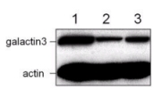
![All lanes : Anti-Galectin 3 antibody [A3A12] (ab2785) at 1/1000 dilutionLane 1 : MCF-7 cell lysateLane 2 : HeLa cell lysateLane 3 : NIH-3T3 cell lysateLysates/proteins at 25 µg/ml per lane.](http://www.bioprodhub.com/system/product_images/ab_products/2/sub_2/22241_ab2785-201418-ab2785wb.jpg) All lanes : Anti-Galectin 3 antibody [A3A12] (ab2785) at 1/1000 dilutionLane 1 : MCF-7 cell lysateLane 2 : HeLa cell lysateLane 3 : NIH-3T3 cell lysateLysates/proteins at 25 µg/ml per lane.
All lanes : Anti-Galectin 3 antibody [A3A12] (ab2785) at 1/1000 dilutionLane 1 : MCF-7 cell lysateLane 2 : HeLa cell lysateLane 3 : NIH-3T3 cell lysateLysates/proteins at 25 µg/ml per lane.
 ICC/IF image of ab2785 stained HeLa cells. The cells were 4% PFA fixed (10 min) and then incubated in 1%BSA / 10% normal goat serum / 0.3M glycine in 0.1% PBS-Tween for 1h to permeabilise the cells and block non-specific protein-protein interactions. The cells were then incubated with the antibody (ab2785, 5µg/ml) overnight at +4°C. The secondary antibody (green) was Alexa Fluor® 488 goat anti-mouse IgG (H+L) used at a 1/1000 dilution for 1h. Alexa Fluor® 594 WGA was used to label plasma membranes (red) at a 1/200 dilution for 1h. DAPI was used to stain the cell nuclei (blue) at a concentration of 1.43µM.
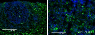 ab2785 staining Galectin 3 in rat spleen tissue by Immunohistochemistry (Frozen sections).Tissue was fixed in paraformaldehyde, blocked with 20% serum for 20 minutes at 24°C, then incubated with ab2785 at a 1/200 dilution for 16 hours at 4°C. The secondary used was an Alexa-Fluor 488 conjugated donkey anti-mouse polyclonal used at a 1/1000 dilution. Counterstained with Hoechst 33258 (blue).See Abreview
Product References
Epigenetic regulation of galectin-3 expression by beta1 integrins promotes cell - Epigenetic regulation of galectin-3 expression by beta1 integrins promotes cell
Margadant C, van den Bout I, van Boxtel AL, Thijssen VL, Sonnenberg A. J Biol Chem. 2012 Dec 28;287(53):44684-93.
Comprehensive analysis of cellular galectin-3 reveals no consistent oncogenic - Comprehensive analysis of cellular galectin-3 reveals no consistent oncogenic
Hann A, Gruner A, Chen Y, Gress TM, Buchholz M. PLoS One. 2011;6(6):e20859.
.
Aberrant epithelial morphology and persistent epidermal growth factor receptor - Aberrant epithelial morphology and persistent epidermal growth factor receptor
Morris ZS, McClatchey AI. Proc Natl Acad Sci U S A. 2009 Jun 16;106(24):9767-72. doi:
Roscovitine reduces neuronal loss, glial activation, and neurologic deficits - Roscovitine reduces neuronal loss, glial activation, and neurologic deficits
Hilton GD, Stoica BA, Byrnes KR, Faden AI. J Cereb Blood Flow Metab. 2008 Nov;28(11):1845-59.
Expression of the transcription factor NFAT2 in human neutrophils: IgE-dependent, - Expression of the transcription factor NFAT2 in human neutrophils: IgE-dependent,
Vega A, Chacon P, Monteseirin J, El Bekay R, Alba G, Martin-Nieto J, Sobrino F. J Cell Sci. 2007 Jul 15;120(Pt 14):2328-37.
Expression of two temporally distinct microglia-related gene clusters after - Expression of two temporally distinct microglia-related gene clusters after
Byrnes KR, Garay J, Di Giovanni S, De Biase A, Knoblach SM, Hoffman EP, Movsesyan V, Faden AI. Glia. 2006 Mar;53(4):420-33.
RNA interference-mediated choline kinase suppression in breast cancer cells - RNA interference-mediated choline kinase suppression in breast cancer cells
Glunde K, Raman V, Mori N, Bhujwalla ZM. Cancer Res. 2005 Dec 1;65(23):11034-43.
Modulation of functional properties of galectin-3 by monoclonal antibodies - Modulation of functional properties of galectin-3 by monoclonal antibodies
Liu FT, Hsu DK, Zuberi RI, Hill PN, Shenhav A, Kuwabara I, Chen SS. Biochemistry. 1996 May 14;35(19):6073-9.