Anti-Galanin antibody - C-terminal
| Name | Anti-Galanin antibody - C-terminal |
|---|---|
| Supplier | Abcam |
| Catalog | ab175487 |
| Prices | $384.00 |
| Sizes | 400 µl |
| Host | Rabbit |
| Clonality | Polyclonal |
| Isotype | IgG |
| Applications | FC WB ICC/IF ICC/IF |
| Species Reactivities | Mouse, Human |
| Antigen | Synthetic peptide within Human Galanin aa 80-109 (C terminal) conjugated to Keyhole Limpet Haemocyanin (KLH) |
| Description | Rabbit Polyclonal |
| Gene | GAL |
| Conjugate | Unconjugated |
| Supplier Page | Shop |
Product images
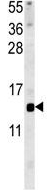 Anti-Galanin antibody - C-terminal (ab175487) at 1/1000 dilution + Mouse liver tissue lysate at 15 µg
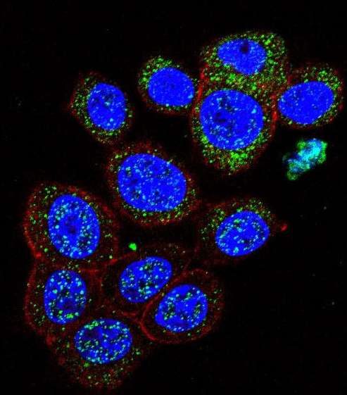 Immunofluorescent analysis of 293 cells labeling Galanin with ab175487 at 1/10 dilution (green). Actin filaments have been labeled with Alexa Fluor 555 phalloidin (red). DAPI was used to stain the cell nuclear (blue).
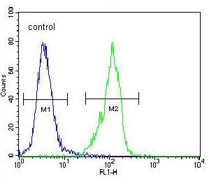 Flow cytometric analysis of 293 cells labeling Galanin with ab175487 antibody at 1/10 dilution (right histogram), compared to a negative control cell (left histogram). FITC-conjugated goat-anti-rabbit secondary antibodies were used for the analysis.