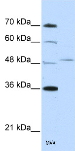
Anti-FOXQ1 antibody (ab51340) at 2.5 µg/ml + Jurkat cell lysate at 10 µg with skim milk/ PBS at 5 %SecondaryHRP conjugated anti-Rabbit IgG at 1/50000 dilution
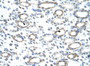
Immunohistochemistry (Formalin/PFA-fixed paraffin-embedded sections) analysis of human kidney tissue labelling FOXQ1 with ab51340 at 4-8µg/ml. Arrows indicate postively labelled epithelial cells of renal tubule. Magnification: 400X.
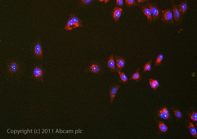
ICC/IF image of ab51340 stained cells. The cells were and then incubated in 1%BSA / 10% normal goat serum / 0.3M glycine in 0.1% PBS-Tween for 1h to permeabilise the cells and block non-specific protein-protein interactions. The cells were then incubated with the antibody (ab51340, 5µg/ml) overnight at +4°C. The secondary antibody (green) was ab96899 Dylight 488 goat anti-rabbit IgG (H+L) used at a 1/250 dilution for 1h. Alexa Fluor® 594 WGA was used to label plasma membranes (red) at a 1/200 dilution for 1h. DAPI was used to stain the cell nuclei (blue) at a concentration of 1.43µM.