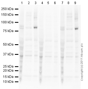
All lanes : Anti-FOXO3A (phospho T32) antibody (ab26649) at 1 µg/mlLane 1 : HT29 (Human colon adenocarcinoma grade II cell line) Whole Cell Lysate (ab3952)Lane 2 : Jurkat (Human T cell lymphoblast-like cell line) Whole Cell Lysate Lane 3 : 293T Cell extract - treated with Calyculin ALane 4 : HT29 (Human colon adenocarcinoma grade II cell line) Whole Cell Lysate (ab3952) with Human FOXO3A (phospho T32) peptide (ab27877) at 1 mg/mlLane 5 : Jurkat (Human T cell lymphoblast-like cell line) Whole Cell Lysate with Human FOXO3A (phospho T32) peptide (ab27877) at 1 mg/mlLane 6 : 293T Cell extract - treated with Calyculin A with Human FOXO3A (phospho T32) peptide (ab27877) at 1 mg/mlLane 7 : HT29 (Human colon adenocarcinoma grade II cell line) Whole Cell Lysate (ab3952) with Human FOXO3A peptide (ab27878) at 1 mg/mlLane 8 : Jurkat (Human T cell lymphoblast-like cell line) Whole Cell Lysate with Human FOXO3A peptide (ab27878) at 1 mg/mlLane 9 : 293T Cell extract - treated with Calyculin A with H

ab26649 specifically recognises the immunizing FOXO3A (phospho T32) peptide in ELISA analysis (orange dotted line), but not the corresponding unmodified peptide (yellow dotted line).

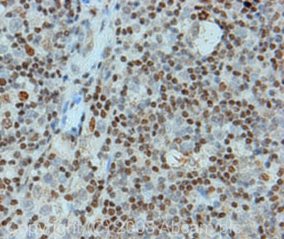
IHC image of FOXO3A staining in human Hodgkin's lymphoma FFPE section, performed on a BondTM system using the standard protocol F. The section was pre-treated using heat mediated antigen retrieval with sodium citrate buffer (pH6, epitope retrieval solution 1) for 20 mins. The section was then incubated with ab26649, 5µg/ml, for 8 mins at room temperature and detected using an HRP conjugated compact polymer system. DAB was used as the chromogen. The section was then counterstained with haematoxylin and mounted with DPX.


