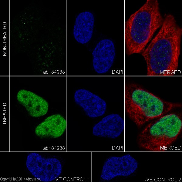
Immunofluorescent analysis of 4% paraformaldehyde-fixed, 0.1% Triton X-100 permeabilized HeLa (Human epithelial cells from cervix adenocarcinoma) cells, untreated or treated with 12-o-tetradecanoyl phorbol 13-acetate (TPA) (200nM) for 4h, labeling Fos B with ab184938 at 1/500 dilution, followed by Goat anti-rabbit IgG (Alexa Fluor® 488) (ab150077) secondary antibody at 1/1000 dilution (green).Confocal image showing nuclear staining on HeLa cells. The expression was increaed after treatment with 12-o-tetradecanoyl phorbol 13-acetate (TPA) (200nM) for 4h. The nuclear counter stain is DAPI (blue). Tubulin is detected with ab7291 (anti-Tubulin mouse mAb) at 1/1000 dilution and ab150120 (AlexaFluor®594 Goat anti-Mouse secondary) at 1/500 dilution (red).The negative controls are as follows:-ve control 1: ab184938 at 1/1000 dilution followed by ab150120 (AlexaFluor®594 Goat anti-Mouse secondary) at 1/500 dilution.-ve control 2: ab7291 (anti-Tubulin mouse mAb) at 1/1000 dilution followed by ab
![All lanes : Anti-Fos B antibody [EPR15905] (ab184938) at 1/10000 dilutionLane 1 : Untreated HeLa (Human epithelial cells from cervix adenocarcinoma) whole cell lysateLane 2 : HeLa whole cell lysate serum starved overnight, then treated with 200nM 12-o-tetradecanoyl phorbol 13-acetate (TPA) for 4hr.Lysates/proteins at 10 µg per lane.SecondaryGoat Anti-Rabbit IgG, (H+L),Peroxidase conjugated at 1/1000 dilution](http://www.bioprodhub.com/system/product_images/ab_products/2/sub_2/20194_ab184938-242386-184938WBa.jpg)
All lanes : Anti-Fos B antibody [EPR15905] (ab184938) at 1/10000 dilutionLane 1 : Untreated HeLa (Human epithelial cells from cervix adenocarcinoma) whole cell lysateLane 2 : HeLa whole cell lysate serum starved overnight, then treated with 200nM 12-o-tetradecanoyl phorbol 13-acetate (TPA) for 4hr.Lysates/proteins at 10 µg per lane.SecondaryGoat Anti-Rabbit IgG, (H+L),Peroxidase conjugated at 1/1000 dilution
![All lanes : Anti-Fos B antibody [EPR15905] (ab184938) at 1/10000 dilutionLane 1 : Untreated NIH/3T3 (Mouse embyro fibroblast cells) whole cell lysateLane 2 : NIH/3T3 whole cell lysate treated with serum, starved overnight, then serum stimulated for 4hrLysates/proteins at 10 µg per lane.SecondaryGoat Anti-Rabbit IgG, (H+L),Peroxidase conjugated at 1/1000 dilution](http://www.bioprodhub.com/system/product_images/ab_products/2/sub_2/20195_ab184938-242385-184938WBb.jpg)
All lanes : Anti-Fos B antibody [EPR15905] (ab184938) at 1/10000 dilutionLane 1 : Untreated NIH/3T3 (Mouse embyro fibroblast cells) whole cell lysateLane 2 : NIH/3T3 whole cell lysate treated with serum, starved overnight, then serum stimulated for 4hrLysates/proteins at 10 µg per lane.SecondaryGoat Anti-Rabbit IgG, (H+L),Peroxidase conjugated at 1/1000 dilution
![All lanes : Anti-Fos B antibody [EPR15905] (ab184938) at 1/10000 dilutionLane 1 : Untreated C6 (Rat glial tumor cells) whole cell lysateLane 2 : C6 whole cell lysate treated with serum, starved overnight, then serum stimulated for 4hrLysates/proteins at 10 µg per lane.SecondaryGoat Anti-Rabbit IgG, (H+L),Peroxidase conjugated at 1/1000 dilution](http://www.bioprodhub.com/system/product_images/ab_products/2/sub_2/20196_ab184938-242384-184938WBc.jpg)
All lanes : Anti-Fos B antibody [EPR15905] (ab184938) at 1/10000 dilutionLane 1 : Untreated C6 (Rat glial tumor cells) whole cell lysateLane 2 : C6 whole cell lysate treated with serum, starved overnight, then serum stimulated for 4hrLysates/proteins at 10 µg per lane.SecondaryGoat Anti-Rabbit IgG, (H+L),Peroxidase conjugated at 1/1000 dilution
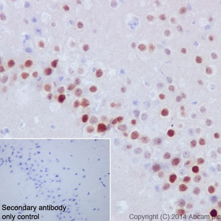
Immunohistochemical analysis of paraffin-embedded mouse hippocampus tissue labeling Fos B with ab184938 at 1/1000 dilution, followed by Goat Anti-Rabbit IgG H&L (HRP) (ab97051) secondary antibody at 1/500 dilution. Nucleus staining on mouse hippocampus is observed. Counter stained with Hematoxylin.Secondary antibody only control: Used PBS instead of primary antibody, secondary antibody is Goat Anti-Rabbit IgG H&L (HRP) (ab97051) at 1/500 dilution.
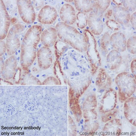
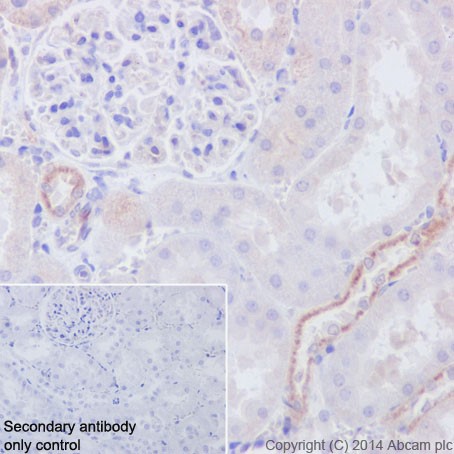
Immunohistochemical analysis of paraffin-embedded mouse kidney tissue labeling Fos B with ab184938 at 1/1000 dilution, followed by Goat Anti-Rabbit IgG H&L (HRP) (ab97051) secondary antibody at 1/500 dilution. Cytoplasm staining on mouse kidney is observed. Counter stained with Hematoxylin.Secondary antibody only control: Used PBS instead of primary antibody, secondary antibody is Goat Anti-Rabbit IgG H&L (HRP) (ab97051) at 1/500 dilution.
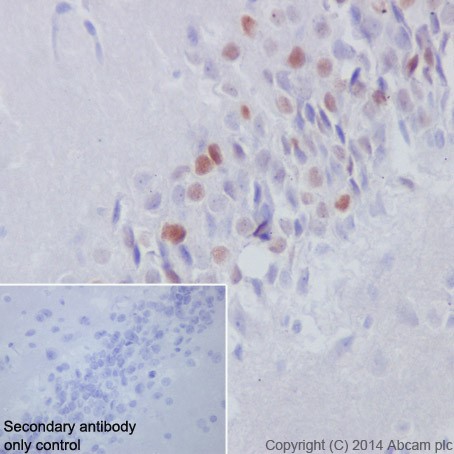
Immunohistochemical analysis of paraffin-embedded rat hippocampus tissue labeling Fos B with ab184938 at 1/1000 dilution, followed by Goat Anti-Rabbit IgG H&L (HRP) (ab97051) secondary antibody at 1/500 dilution. Nucleus staining on rat hippocampus is observed. Counter stained with Hematoxylin.Secondary antibody only control: Used PBS instead of primary antibody, secondary antibody is Goat Anti-Rabbit IgG H&L (HRP) (ab97051) at 1/500 dilution.
Immunohistochemical analysis of paraffin-embedded rat kidney tissue labeling Fos B with ab184938 at 1/1000 dilution, followed by Goat Anti-Rabbit IgG H&L (HRP) (ab97051) secondary antibody at 1/500 dilution. Cytoplasm staining on rat kidney is observed. Counter stained with Hematoxylin.Secondary antibody only control: Used PBS instead of primary antibody, secondary antibody is Goat Anti-Rabbit IgG H&L (HRP) (ab97051) at 1/500 dilution.
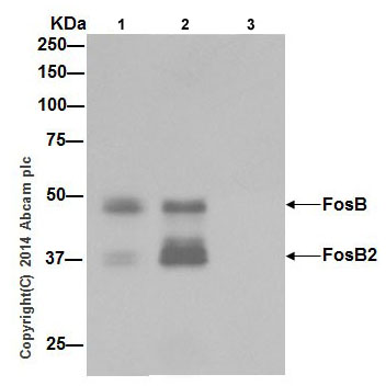
Fos B was immunoprecipitated from 1mg of HeLa (Human epithelial cells from cervix adenocarcinoma) whole cell lysate serum starved overnight, then treated with 200nM 12-o-tetradecanoyl phorbol 13-acetate (TPA) for 4hr using ab184938 at 1/50 dilution.Western blot was performed from the immunoprecipitate using ab184938 at 1/1000 dilution. Anti-Rabbit IgG (HRP), specific to the non-reduced form of IgG, was used as secondary antibody at 1/1500 dilution.Lane 1: HeLa whole cell lysate serum starved overnight, then treated with 200nM 12-o-tetradecanoyl phorbol 13-acetate (TPA) for 4hr, 10 µg (Input).Lane 2: ab184938 IP in HeLa whole cell lysate serum starved overnight, then treated with 200nM 12-o-tetradecanoyl phorbol 13-acetate (TPA) for 4hr.Lane 3: Rabbit monoclonal IgG (ab172730) instead of ab184938 in HeLa whole cell lysate serum starved overnight, then treated with 200nM 12-o-tetradecanoyl phorbol 13-acetate (TPA) for 4hr.Blocking and dilution buffer and concentration: 5% NFDM/TBST.