Anti-FKBP12 antibody
| Name | Anti-FKBP12 antibody |
|---|---|
| Supplier | Abcam |
| Catalog | ab2918 |
| Prices | $400.00 |
| Sizes | 100 µl |
| Host | Rabbit |
| Clonality | Polyclonal |
| Isotype | IgG |
| Applications | IHC-P ICC/IF ICC/IF IP WB IHC-F |
| Species Reactivities | Mouse, Rat, Dog, Human, Pig, Rabbit, Bovine, Xenopus |
| Antigen | Synthetic peptide corresponding to Human FKBP12 aa 1-13 (N terminal) |
| Description | Rabbit Polyclonal |
| Gene | FKBP1A |
| Conjugate | Unconjugated |
| Supplier Page | Shop |
Product images
 ab2918 at 1/1000 staining FKBP12 from human penis tissue by IHC-P. The tissue was paraformaldehyde fixed and the slides were incubated in citrate buffer for antigen retrieval and heated for 20 minutes. The tissue was incubated with ab2918 for 24 hours. The secondary antibody used was part of the DAKO ENVISON System. The picture depicts staining of nerve fibers within corporal tissue of the human penis.See Abreview
 All lanes : Anti-FKBP12 antibody (ab2918) at 1/1000 dilutionLane 1 : Whole tissue lysate, dog left ventricle (heart failure)Lane 2 : Whole tissue lysate, dog left ventricle (heart failure)Lane 3 : Whole tissue lysate, dog left ventricle (heart failure)Lane 4 : Whole tissue lysate, dog left ventricle (heart failure)Lane 5 : Whole tissue lysate, dog left ventricle (heart failure)Lane 6 : Whole tissue lysate, dog left ventricle (normal)Lane 7 : Whole tissue lysate, dog left ventricle (normal)Lane 8 : Whole tissue lysate, dog left ventricle (normal)Lane 9 : Whole tissue lysate, dog left ventricle (normal)Lysates/proteins at 20 µg per lane.SecondaryHRP conjugated donkey polyclonal antibodydeveloped using the ECL techniquePerformed under reducing conditions.Observed band size : 12 kDa (why is the actual band size different from the predicted?)Exposure time : 2 minutesThis image is courtesy of an Abreview submitted by Dr sudhish mishraSee Abreview
 ab2918 at 1/1000 staining mouse heart tissue sections by IHC-P. The tissue was paraformaldehyde fixed, blocked and then a citrate buffer / heat mediated antigen retireival step was performed. The tissue was incubated with the antibody for 24 hours. An HRP conjugated rabbit polyclonal antibody was used as the secondary.See Abreview
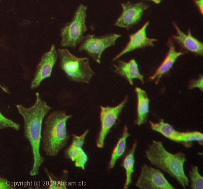 ICC/IF image of ab2918 stained HepG2 cells. The cells were 4% formaldehyde fixed (10 min) and then incubated in 1%BSA / 10% normal goat serum / 0.3M glycine in 0.1% PBS-Tween for 1h to permeabilise the cells and block non-specific protein-protein interactions. The cells were then incubated with the antibody (ab2918, 1µg/ml) overnight at +4°C. The secondary antibody (green) was Alexa Fluor® 488 goat anti-rabbit IgG (H+L) used at a 1/1000 dilution for 1h. Alexa Fluor® 594 WGA was used to label plasma membranes (red) at a 1/200 dilution for 1h. DAPI was used to stain the cell nuclei (blue) at a concentration of 1.43µM.
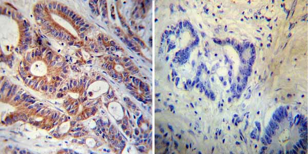 Immunohistochemistry was performed on both normal and cancer biopsies of deparaffinized human colon carcinoma tissue. To expose target proteins, heat induced antigen retrieval was performed using 10mM sodium citrate (pH 6.0) buffer, microwaved for 8-15 minutes. Following antigen retrieval tissues were blocked in 3% BSA-PBS for 30 minutes at room temperature. Tissues were then probed at a dilution of 1/200 with a rabbit polyclonal antibody recognizing FKBP12 (ab2918) or without primary antibody (negative control) overnight at 4°C in a humidified chamber. Tissues were washed extensively with PBST and endogenous peroxidase activity was quenched with a peroxidase suppressor. Detection was performed using a biotin-conjugated secondary antibody and SA-HRP, followed by colorimetric detection using DAB. Tissues were counterstained with hematoxylin and prepped for mounting.
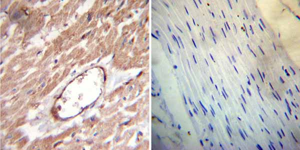 Immunohistochemistry was performed on both normal and cancer biopsies of deparaffinized human heart tissue. To expose target proteins, heat induced antigen retrieval was performed using 10mM sodium citrate (pH 6.0) buffer, microwaved for 8-15 minutes. Following antigen retrieval tissues were blocked in 3% BSA-PBS for 30 minutes at room temperature. Tissues were then probed at a dilution of 1/20 with a rabbit polyclonal antibody recognizing FKBP12 (ab2918) or without primary antibody (negative control) overnight at 4°C in a humidified chamber. Tissues were washed extensively with PBST and endogenous peroxidase activity was quenched with a peroxidase suppressor. Detection was performed using a biotin-conjugated secondary antibody and SA-HRP, followed by colorimetric detection using DAB. Tissues were counterstained with hematoxylin and prepped for mounting.
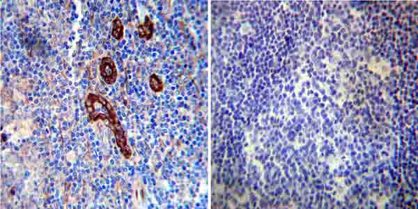 Immunohistochemistry was performed on both normal and cancer biopsies of deparaffinized human tonsil tissue. To expose target proteins, heat induced antigen retrieval was performed using 10mM sodium citrate (pH 6.0) buffer, microwaved for 8-15 minutes. Following antigen retrieval tissues were blocked in 3% BSA-PBS for 30 minutes at room temperature. Tissues were then probed at a dilution of 1/50 with a rabbit polyclonal antibody recognizing FKBP12 (ab2918) or without primary antibody (negative control) overnight at 4°C in a humidified chamber. Tissues were washed extensively with PBST and endogenous peroxidase activity was quenched with a peroxidase suppressor. Detection was performed using a biotin-conjugated secondary antibody and SA-HRP, followed by colorimetric detection using DAB. Tissues were counterstained with hematoxylin and prepped for mounting.
Product References
Recruitment of Mad1 to metaphase kinetochores is sufficient to reactivate the - Recruitment of Mad1 to metaphase kinetochores is sufficient to reactivate the
Ballister ER, Riegman M, Lampson MA. J Cell Biol. 2014 Mar 17;204(6):901-8.
Forced dimerization increases the activity of DeltaEGFR/EGFRvIII and enhances its - Forced dimerization increases the activity of DeltaEGFR/EGFRvIII and enhances its
Hwang Y, Chumbalkar V, Latha K, Bogler O. Mol Cancer Res. 2011 Sep;9(9):1199-208.
Development and application of in vivo molecular traps reveals that dynein light - Development and application of in vivo molecular traps reveals that dynein light
Varma D, Dawn A, Ghosh-Roy A, Weil SJ, Ori-McKenney KM, Zhao Y, Keen J, Vallee RB, Williams JC. Proc Natl Acad Sci U S A. 2010 Feb 23;107(8):3493-8. doi:
A molecularly engineered split reporter for imaging protein-protein interactions - A molecularly engineered split reporter for imaging protein-protein interactions
Massoud TF, Paulmurugan R, Gambhir SS. Nat Med. 2010 Aug;16(8):921-6.
Generation of Epstein-Barr virus-specific cytotoxic T lymphocytes resistant to - Generation of Epstein-Barr virus-specific cytotoxic T lymphocytes resistant to
De Angelis B, Dotti G, Quintarelli C, Huye LE, Zhang L, Zhang M, Pane F, Heslop HE, Brenner MK, Rooney CM, Savoldo B. Blood. 2009 Nov 26;114(23):4784-91.
Selective clearance of macrophages in atherosclerotic plaques by autophagy. - Selective clearance of macrophages in atherosclerotic plaques by autophagy.
Verheye S, Martinet W, Kockx MM, Knaapen MW, Salu K, Timmermans JP, Ellis JT, Kilpatrick DL, De Meyer GR. J Am Coll Cardiol. 2007 Feb 13;49(6):706-15. Epub 2007 Jan 26.
Expression profiling of laser-microdissected intrapulmonary arteries in - Expression profiling of laser-microdissected intrapulmonary arteries in
Kwapiszewska G, Wilhelm J, Wolff S, Laumanns I, Koenig IR, Ziegler A, Seeger W, Bohle RM, Weissmann N, Fink L. Respir Res. 2005 Sep 19;6:109.