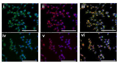
ab80264 staining FJX1 by ICC/IF (Immunocytochemistry/immunofluorescence) in HEK293T cells stably expressing MYC-tagged FJX1 dual stained for FJX1 (i, green) and MYC (ii, red) or FJX1 (iv, green) and the Golgi marker, GM130 (v, red). Nuclei were stained with 4',6-diamidino-2-phenylindole (blue). Respective merged images are shown (iii, vi). Scale bar = 100 μmCells were fixed with 4% paraformaldehyde, blocked with 3% BSA and incubated with primary antibody and an Alexa Fluor® was used as the secondary antibody.