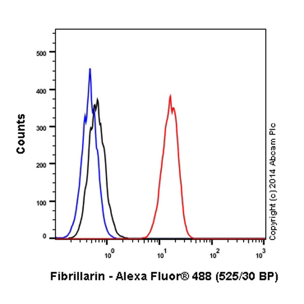
Overlay histogram showing HeLa cells stained with ab184817 (red line). The cells were fixed with 80% methanol (5 min) and then permeabilized with 0.1% PBS-Tween for 20 min. The cells were then incubated in 1x PBS / 10% normal goat serum / 0.3M glycine to block non-specific protein-protein interactions followed by the antibody (ab184817, 1/500 dilution) for 30 min at 22°C. Isotype control antibody (black line) was rabbit IgG (monoclonal) Alexa Fluor® 488 used at the same concentration and conditions as the primary antibody. Unlabelled sample (blue line) was also used as a control.Acquisition of >5,000 events were collected using a 20mW Argon ion laser (488nm) and 525/30 bandpass filter.This antibody gave a positive signal in HeLa fixed with 4% formaldehyde (10 min)/permeabilized with 0.1% PBS-Tween for 20 min used under the same conditions.
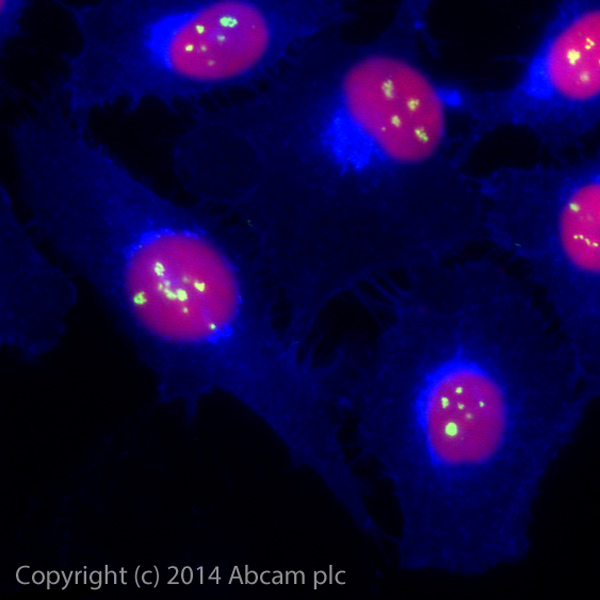
ab184817 staining Fibrillarin in HeLa cells. The cells were fixed with 100% methanol (5min) and then blocked in 1% BSA/10% normal goat serum/0.3M glycine in 0.1% PBS-Tween for 1h. The cells were then incubated with ab184817 at a working diltuion of 1 in 100 overnight at +4°C (shown in green). AlexaFluor® 350 WGA was used at a 1/200 dilution and incubated for 1h with the cells, to label plasma membranes (shown in blue). Nuclear DNA was labelled in red with 1.25 μM DRAQ5™ (ab108410).