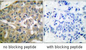
Immunohistochemical analysis of paraffin-embedded human breast carcinoma tissue using ab53074 at 1/50 dilution, with and without peptide immunogen.
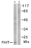
All lanes : Anti-FHIT antibody (ab53074) at 1/500 dilutionLane 1 : A549 cell extract with no immunizing peptideLane 2 : A549 cell extract with immunizing peptide
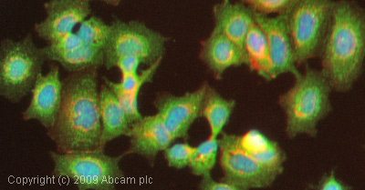
ICC/IF image of ab53074 stained HepG2 cells. The cells were 4% PFA fixed (10 min) and then incubated in 1%BSA / 10% normal goat serum / 0.3M glycine in 0.1% PBS-Tween for 1h to permeabilise the cells and block non-specific protein-protein interactions. The cells were then incubated with the antibody (ab53074, 1µg/ml) overnight at +4°C. The secondary antibody (green) was Alexa Fluor® 488 goat anti-rabbit IgG (H+L) used at a 1/1000 dilution for 1h. Alexa Fluor® 594 WGA was used to label plasma membranes (red) at a 1/200 dilution for 1h. DAPI was used to stain the cell nuclei (blue) at a concentration of 1.43µM.