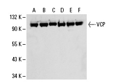
VCP (5): sc-57492. Western blot analysis of VCP expression in NIH/3T3 (A), MDA-MB-231 (B), HeLa (C), MCF7 (D), A-431 (E) and PC-12 (F) whole cell lysates.
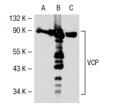
VCP (5): sc-57492. Western blot analysis of VCP expression in non-transfected 293T: sc-117752 (A), human VCP transfected 293T: sc-112114 (B) and NIH/3T3 (C) whole cell lysates.
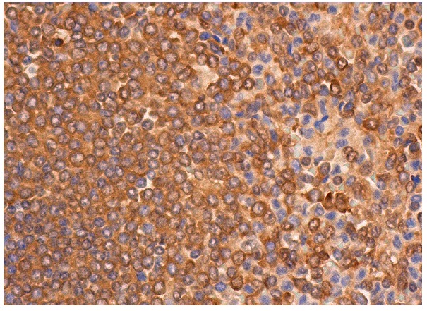
VCP (5): sc-57492. Immunoperoxidase staining of formalin fixed, paraffin-embedded human spleen tissue showing cytoplasmic staining of cells in white pulp and cells in red pulp.