![All lanes : Anti-FGFR2 antibody [3F8] (ab119237) at 1/500 dilutionLane 1 : Lysate of HEK293T cells transfected with pCMV6-ENTRY control cDNALane 2 : Lysate of HEK293T cells transfected with pCMV6-ENTRY FGFR2 cDNA.Lysates/proteins at 5 µg per lane.](http://www.bioprodhub.com/system/product_images/ab_products/2/sub_2/18831_FGFR2-Primary-antibodies-ab119237-4.jpg)
All lanes : Anti-FGFR2 antibody [3F8] (ab119237) at 1/500 dilutionLane 1 : Lysate of HEK293T cells transfected with pCMV6-ENTRY control cDNALane 2 : Lysate of HEK293T cells transfected with pCMV6-ENTRY FGFR2 cDNA.Lysates/proteins at 5 µg per lane.
ab119237, at 1/150, staining FGFR2 by immunohistochemistry of paraffin-embedded Human breast tissue
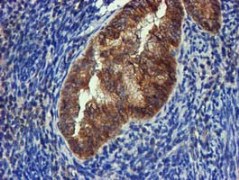
ab119237, at 1/150, staining FGFR2 by immunohistochemistry of paraffin-embedded adenocarcinoma of Human endometrium tissue

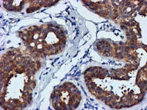
ab119237, at 1/150, staining FGFR2 by immunohistochemistry of paraffin-embedded Human kidney tissue
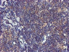
ab119237, at 1/150, staining FGFR2 by immunohistochemistry of paraffin-embedded Human lymphoma tissue
ab119237, at 1/150, staining FGFR2 by immunohistochemistry of paraffin-embedded Human pancreas tissue
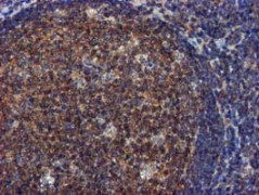
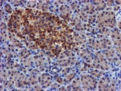
ab119237, at 1/150, staining FGFR2 by immunohistochemistry of paraffin-embedded Human tonsil tissue
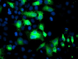
ab119237, at 1/100, staining COS7 cells transiently transfected with pCMV6-ENTRY FGFR2 cDNA, by immunofluorescence
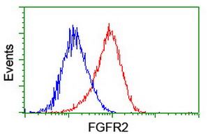
Flow cytometric analysis of HeLa cells, using ab119237 (red),at 1/100, compared to a nonspecific negative control antibody(blue)
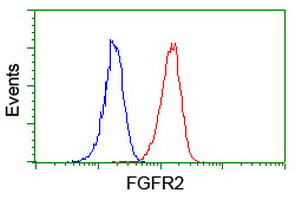
Flow cytometric analysis of Jurkat cells, using ab119237 (red), at 1/100, compared to a nonspecific negative control antibody (blue)