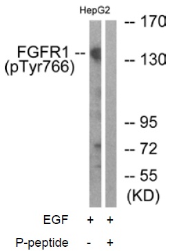
All lanes : Anti-FGFR1 (phospho Y766) antibody (ab59180)Lane 1 : EGF-treated HepG2 cell extractLane 2 : EGF-treated HepG2 cell extract with blocking phosphopeptide

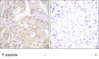
Immunohistochemistry analysis of paraffin-embedded human breast carcinoma tissue using ab59180 with and without the addition of the synthetic phosphopeptide derived from human FGFR1 around the phosphorylation site of tyrosine 766.

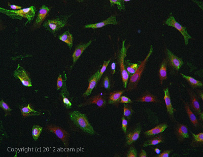
ab59180 stained SKNSH cells. The cells were 4% formaldehyde fixed (10 min) and then incubated in 1%BSA / 10% normal goat serum / 0.3M glycine in 0.1% PBS-Tween for 1h to permeabilise the cells and block non-specific protein-protein interactions. The cells were then incubated with the antibody ab59180 at 5µg/ml overnight at +4°C. The secondary antibody (green) was DyLight® 488 goat anti- rabbit (ab96899) IgG (H+L) used at a 1/1000 dilution for 1h. Alexa Fluor® 594 WGA was used to label plasma membranes (red) at a 1/200 dilution for 1h. DAPI was used to stain the cell nuclei (blue) at a concentration of 1.43µM.


