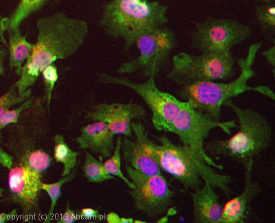
ICC/IF image of ab9588 stained HepG2 cells. The cells were 4% formaldehyde fixed (10 min) and then incubated in 1%BSA / 10% normal goat serum / 0.3M glycine in 0.1% PBS-Tween for 1h to permeabilise the cells and block non-specific protein-protein interactions. The cells were then incubated with the antibody (ab9588, 1µg/ml) overnight at +4°C. The secondary antibody (green) was ab96899, DyLight® 488 goat anti-rabbit IgG (H+L) used at a 1/250 dilution for 1h. Alexa Fluor® 594 WGA was used to label plasma membranes (red) at a 1/200 dilution for 1h. DAPI was used to stain the cell nuclei (blue) at a concentration of 1.43µM.
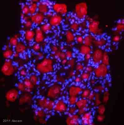
ab9588 staining FGF1 in rat dorsal root ganglia tissue by Immunohistochemistry (Frozen sections).Tissue was fixed in paraformaldehyde and permeabilized using 0.25% Triton X-100. Samples were then blocked 0.25% BSA for 1 hour at 25°C, then incubated with ab9588 at a 1/200 dilution for 16 hours at 4°C. The secondary used was a donkey anti-rabbit polyclonal conjugated to rhodamine, 1/500 dilution.See Abreview