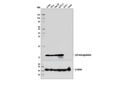
Western blot analysis of extracts from various cell lines using β2-microglobulin (D8P1H) Rabbit mAb (upper) and β-Actin (DA8) Rabbit mAb #8457 (lower). DLD-1 and Daudi cell lines are negative for β2-microglobulin due to genomic deletions at the β2-microglobulin locus.
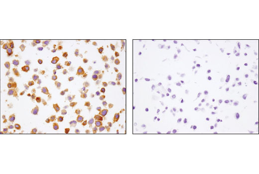
Immunohistochemical analysis of paraffin-embedded HeLa (left) and DLD-1 (right) cell pellets using β2-microglobulin (D8P1H) Rabbit mAb.
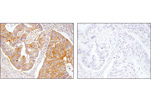
Immunohistochemical analysis of paraffin-embedded colon carcinoma using β2-microglobulin (D8P1H) Rabbit mAb in the presence of control peptide (left) or antigen-specific peptide (right).
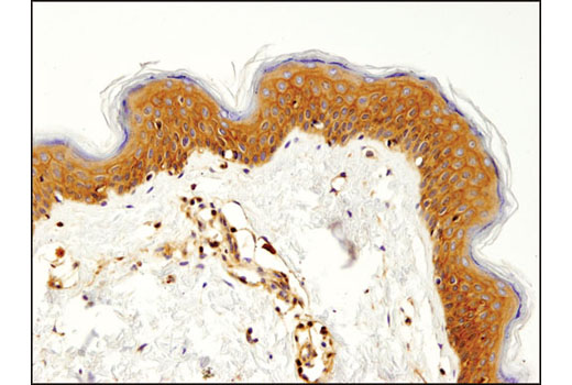
Immunohistochemical analysis of paraffin-embedded human skin using β2-microglobulin (D8P1H) Rabbit mAb.
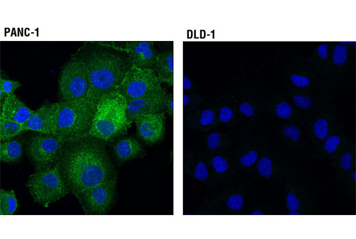
Confocal immunofluorescent analysis of PANC-1 (positive, left) and DLD-1 (negative, right) cells, using β2-microglobulin (D8P1H) Rabbit mAb (green). Blue pseudocolor= DRAQ5 ® #4084 (fluorescent DNA dye).
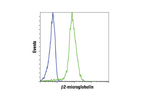
Flow cytometric analysis of DLD-1 cells (blue) and HeLa cells (green) using β2-microglobulin (D8P1H) Rabbit mAb. Anti-rabbit IgG (H+L), F(ab') 2 Fragment (Alexa Fluor ® 647 Conjugate) #4414 was used as a secondary antibody.