Anti-FBXL11 antibody - ChIP Grade
| Name | Anti-FBXL11 antibody - ChIP Grade |
|---|---|
| Supplier | Abcam |
| Catalog | ab31739 |
| Prices | $387.00 |
| Sizes | 100 µg |
| Host | Rabbit |
| Clonality | Polyclonal |
| Isotype | IgG |
| Applications | WB ICC/IF ICC/IF IHC-P ChIP IP |
| Species Reactivities | Human, Mouse |
| Antigen | Synthetic peptide conjugated to KLH derived from within residues 1 - 100 of Human FBXL11 |
| Description | Rabbit Polyclonal |
| Gene | KDM2A |
| Conjugate | Unconjugated |
| Supplier Page | Shop |
Product images
 All lanes : Anti-FBXL11 antibody - ChIP Grade (ab31739) at 1 µg/mlLane 1 : HeLa (Human epithelial carcinoma cell line) Whole Cell Lysate Lane 2 : Jurkat (Human T cell lymphoblast-like cell line) Whole Cell Lysate (ab7899)Lane 3 : HeLa (Human epithelial carcinoma cell line) Nuclear LysateLysates/proteins at 20 µg per lane.SecondaryGoat polyclonal to Rabbit IgG (Alexa Fluor® 680) at 1/10000 dilutionPerformed under reducing conditions.
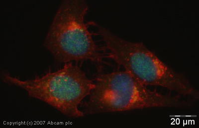 ICC/IF image of ab31739 stained human HeLa cells. The cells were 4% PFA fixed (10 min), permabilised in TBS-T (20 min) and incubated with the antibody (ab31739, 1µg/ml) for 1h at room temperature. 1%BSA / 10% normal goat serum / 0.3M glycine was used to quench autofluorescence and block non-specific protein-protein interactions. The secondary antibody (green) was Alexa Fluor® 488 goat anti-rabbit IgG (H+L) used at a 1/1000 dilution for 1h. Alexa Fluor® 594 WGA was used to label plasma membranes (red). DAPI was used to stain the cell nuclei (blue).
 Image courtesy of Human Protein Atlasab31739 staining KDM2A in human skin, showing a distinct nuclear staining pattern in the epidermis. Paraffin-embedded tissue sections (4µm) were incubated with ab31739 (1/75 dilution) for 30 minutes at room temperature. Antigen retrieval by heat induction in citrate buffer pH 6.ab31739 was tested in a tissue microarray (TMA) containing a wide range of normal and cancer tissues as well as a cell microarray consisting of a range of commonly used, well characterised human cell lines. Further results for this antibody can be found at www.proteinatlas.org
Product References
Depletion of histone demethylase KDM2A enhanced the adipogenic and chondrogenic - Depletion of histone demethylase KDM2A enhanced the adipogenic and chondrogenic
Dong R, Yao R, Du J, Wang S, Fan Z. Exp Cell Res. 2013 Nov 1;319(18):2874-82.
Nucleosome-interacting proteins regulated by DNA and histone methylation. - Nucleosome-interacting proteins regulated by DNA and histone methylation.
Bartke T, Vermeulen M, Xhemalce B, Robson SC, Mann M, Kouzarides T. Cell. 2010 Oct 29;143(3):470-84.