![All lanes : Anti-EXOSC1 antibody [EPR13525] (ab181167) at 1/2000 dilutionLane 1 : 293T lysateLane 2 : Jurkat lysateLane 3 : HepG2 lysateLysates/proteins at 20 µg per lane.SecondaryGoat Anti-Rabbit IgG, (H+L), Peroxidase conjugated at 1/1000 dilution](http://www.bioprodhub.com/system/product_images/ab_products/2/sub_2/16034_ab181167-214634-ab181167wb1.jpg)
All lanes : Anti-EXOSC1 antibody [EPR13525] (ab181167) at 1/2000 dilutionLane 1 : 293T lysateLane 2 : Jurkat lysateLane 3 : HepG2 lysateLysates/proteins at 20 µg per lane.SecondaryGoat Anti-Rabbit IgG, (H+L), Peroxidase conjugated at 1/1000 dilution
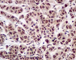
Immunohistochemical analysis of paraffin embedded Human breast carcinoma tissue labeling EXOSC1 with ab181167 at a 1/100 dilution. Prediluted HRP Polymer for Rabbit IgG secondary used. Counterstained with Hematoxylin.
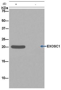
Detection of Human EXOSC1 by Western Blot of Immunoprecipitate. 293T whole cell lysate (lane 1) and a negative control (lane 2) loaded, ab181167 used for blotting immunoprecipitated EXOSC1 at a 1/50 dilution. Anti-Rabbit IgG (HRP), specific to the non-reduced form of IgG secondary used at a 1/1500 dilution.
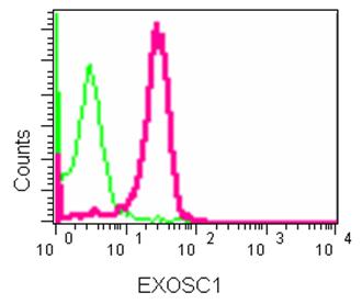
Flow cytometry analysis of K562 cells using ab181167 at a 1/90 dilution (red) or a Rabbit monoclonal IgG (negative) (green). Goat anti rabbit IgG (FITC) secondary used at a 1/150 dilution.
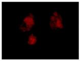
Immunofluorescence analysis of HepG2 cells (fixative 4% paraformaldehyde) labeling EXOSC1 with ab181167 at a 1/250 dilution. Goat anti rabbit IgG (Alexa Fluor® 555) secondary used at a 1/200 diution.