![Anti-ERK1 + ERK2 antibody [EPR17526] (ab184699) at 1/10000 dilution + Human ERK1 full length protein (ab43623) at 0.01 µgSecondaryGoat Anti-Rabbit IgG, (H+L),Peroxidase conjugated at 1/1000 dilution](http://www.bioprodhub.com/system/product_images/ab_products/2/sub_2/15063_ab184699-239201-WB-1.jpg)
Anti-ERK1 + ERK2 antibody [EPR17526] (ab184699) at 1/10000 dilution + Human ERK1 full length protein (ab43623) at 0.01 µgSecondaryGoat Anti-Rabbit IgG, (H+L),Peroxidase conjugated at 1/1000 dilution
![Anti-ERK1 + ERK2 antibody [EPR17526] (ab184699) at 1/10000 dilution + Human ERK2 full length protein (ab43625) at 0.01 µgSecondaryGoat Anti-Rabbit IgG, (H+L),Peroxidase conjugated at 1/1000 dilution](http://www.bioprodhub.com/system/product_images/ab_products/2/sub_2/15064_ab184699-239200-WB-2.jpg)
Anti-ERK1 + ERK2 antibody [EPR17526] (ab184699) at 1/10000 dilution + Human ERK2 full length protein (ab43625) at 0.01 µgSecondaryGoat Anti-Rabbit IgG, (H+L),Peroxidase conjugated at 1/1000 dilution
![All lanes : Anti-ERK1 + ERK2 antibody [EPR17526] (ab184699) at 1/50000 dilutionLane 1 : A431 (Human epidermoid carcinoma) whole cell lysatesLane 2 : Jurkat (Human T cell leukemia cells from peripheral blood) whole cell lysatesLane 3 : HeLa (Human epithelial cells from cervix adenocarcinoma) whole cell lysatesLane 4 : HepG2 (Human liver hepatocellular carcinoma) whole cell lysatesLane 5 : Human fetal brain lysatesLane 6 : Human fetal heart lysatesLane 7 : Human fetal kidney lysatesLysates/proteins at 20 µg per lane.SecondaryGoat Anti-Rabbit IgG, (H+L),Peroxidase conjugated at 1/1000 dilution](http://www.bioprodhub.com/system/product_images/ab_products/2/sub_2/15065_ab184699-239199-WB-3.jpg)
All lanes : Anti-ERK1 + ERK2 antibody [EPR17526] (ab184699) at 1/50000 dilutionLane 1 : A431 (Human epidermoid carcinoma) whole cell lysatesLane 2 : Jurkat (Human T cell leukemia cells from peripheral blood) whole cell lysatesLane 3 : HeLa (Human epithelial cells from cervix adenocarcinoma) whole cell lysatesLane 4 : HepG2 (Human liver hepatocellular carcinoma) whole cell lysatesLane 5 : Human fetal brain lysatesLane 6 : Human fetal heart lysatesLane 7 : Human fetal kidney lysatesLysates/proteins at 20 µg per lane.SecondaryGoat Anti-Rabbit IgG, (H+L),Peroxidase conjugated at 1/1000 dilution
![All lanes : Anti-ERK1 + ERK2 antibody [EPR17526] (ab184699) at 1/10000 dilutionLane 1 : Human fetal brain lysatesLane 2 : Human fetal heart lysatesLysates/proteins at 10 µg per lane.SecondaryAnti-Rabbit IgG (HRP), specific to the non-reduced form of IgG at 1/1000 dilution](http://www.bioprodhub.com/system/product_images/ab_products/2/sub_2/15066_ab184699-239198-WB-4.jpg)
All lanes : Anti-ERK1 + ERK2 antibody [EPR17526] (ab184699) at 1/10000 dilutionLane 1 : Human fetal brain lysatesLane 2 : Human fetal heart lysatesLysates/proteins at 10 µg per lane.SecondaryAnti-Rabbit IgG (HRP), specific to the non-reduced form of IgG at 1/1000 dilution
![All lanes : Anti-ERK1 + ERK2 antibody [EPR17526] (ab184699) at 1/10000 dilutionLane 1 : Mouse brain lysatesLane 2 : Mouse heart lysatesLane 3 : Mouse kidney lysatesLane 4 : Mouse spleen lysatesLane 5 : Rat brain lysatesLane 6 : Rat heart lysatesLane 7 : Rat kidney lysatesLane 8 : Rat spleen lysatesLane 9 : C6 (Rat glial tumor cells) whole cell lysatesLane 10 : RAW 264.7 (Mouse macrophage cells transformed with Abelson murine leukemia virus) whole cell lysatesLane 11 : PC-12 (Rat adrenal gland pheochromocytoma) whole cell lysatesLane 12 : NIH/3T3 (Mouse embyro fibroblast cells) whole cell lysatesLysates/proteins at 10 µg per lane.SecondaryGoat Anti-Rabbit IgG, (H+L),Peroxidase conjugated at 1/1000 dilution](http://www.bioprodhub.com/system/product_images/ab_products/2/sub_2/15067_ab184699-239197-WB-5.jpg)
All lanes : Anti-ERK1 + ERK2 antibody [EPR17526] (ab184699) at 1/10000 dilutionLane 1 : Mouse brain lysatesLane 2 : Mouse heart lysatesLane 3 : Mouse kidney lysatesLane 4 : Mouse spleen lysatesLane 5 : Rat brain lysatesLane 6 : Rat heart lysatesLane 7 : Rat kidney lysatesLane 8 : Rat spleen lysatesLane 9 : C6 (Rat glial tumor cells) whole cell lysatesLane 10 : RAW 264.7 (Mouse macrophage cells transformed with Abelson murine leukemia virus) whole cell lysatesLane 11 : PC-12 (Rat adrenal gland pheochromocytoma) whole cell lysatesLane 12 : NIH/3T3 (Mouse embyro fibroblast cells) whole cell lysatesLysates/proteins at 10 µg per lane.SecondaryGoat Anti-Rabbit IgG, (H+L),Peroxidase conjugated at 1/1000 dilution
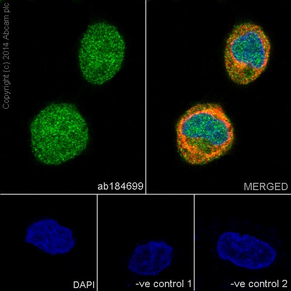
Immunofluorescent analysis of 4% paraformaldehyde-fixed, 0.1% Triton X-100 permeabilized HeLa (Human epithelial cells from cervix adenocarcinoma) cells labeling ERK1 + ERK2 with ab184699 at 1/250 dilution, followed by Goat anti-rabbit IgG (Alexa Fluor® 488) (ab150077) secondary antibody at 1/500 dilution (green). Confocal image showing both nuclear and cytoplasmic staining on HeLa cell line. The nuclear counter stain is DAPI (blue). Tubulin is detected with ab7291 (anti-Tubulin mouse mAb) at 1/1000 dilution and ab150120 (AlexaFluor®594 Goat anti-Mouse secondary) at 1/500 dilution (red).The negative controls are as follows:-ve control 1: ab184699 at 1/250 dilution followed by ab150120 (AlexaFluor®594 Goat anti-Mouse secondary) at 1/500 dilution.-ve control 2: ab7291 (anti-Tubulin mouse mAb) at 1/1000 dilution followed by ab150077 (Alexa Fluor®488 Goat Anti-Rabbit IgG H&L) at 1/500 dilution.
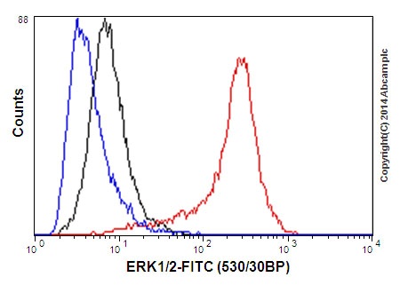
Flow cytometric analysis of A431 (Human epidermoid carcinoma) cells labeling ERK1 + ERK2 with ab184699 at 1/440 dilution (red) compared with a rabbit monoclonal IgG isotype control (black) and an unlabelled control (cells without incubation with primary antibody and secondary antibody; blue). Goat anti rabbit IgG (FITC) at 1/150 dilution was used as the secondary antibody.
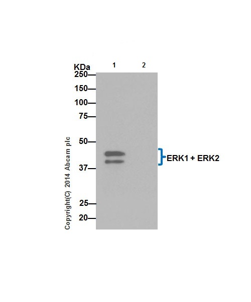
ERK1 + ERK2 were immunoprecipitated from 1mg of PC-12 (Rat adrenal gland pheochromocytoma) whole cell extract with ab184699 at 1/70 dilution. Western blot was performed from the immunoprecipitate using ab184699 at 1/5000 dilution. Anti-Rabbit IgG (HRP), specific to the non-reduced form of IgG, was used as secondary antibody at 1/1500 dilution.Lane 1: PC-12 whole cell extract. Lane 2: PBS instead of PC-12 whole cell extract.Blocking and dilution buffer and concentration: 5% NFDM/TBST.44kDa band represents ERK1. 42kDa band represents ERK2.