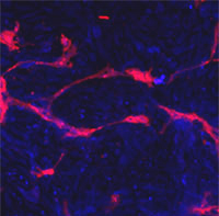
Immunofluorescent staining of methanol/acetone fixed human stem cells for Eph4A (blue) and endothelial Lectin(red). Data kindly provided by Dr. Weis from Cheresh Lab, UCSD.

ab5389 EphA4 antibody immunostaining cortical neurons (image taken at X2; cytoplasmic staining).Bar=20 microns.Protocol: ab5389 incubated overnight at room temperature at a dilution of 1/100-1/300 on free floating perfusion fixed coronal sections of brain. Immunostaining visualised by direct fluorescence (Alexa Fluor® 488, 1/1000).

ab5389 EHpA4 antibody immunostaining hippocampal neurons ( X2 magnification; cytoplasmic staining). Bar: 20 microns.Protocol: ab5389 incubated overnight at room temperature at a dilution of 1/100-1/300 on free floating perfusion fixed coronal sections of brain. Immunostaining visualised by direct fluorescence (Alexa Fluor® 488, 1/1000).

ab5389 staining Eph receptor A4 in human breast carcinoma (BC) tissue by Immunohistochemistry (Formalin/PFA-fixed paraffin-embedded sections). ab5389 was peroxidase-conjugated to the secondary antibody, followed by AEC staining.

Anti-Eph receptor A4 antibody (ab5389) at 1/100 dilution + HeLa cell lysateSecondaryHRP-conjugated anti-rabbit IgG

All lanes : Anti-Eph receptor A4 antibody (ab5389) at 1/1000 dilutionLane 1 : 293 cell lysate - nontransfectedLane 2 : 293 cell lysate - transfected with EphA4Lysates/proteins at 2 µg per lane.

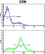
Flow cytometry analysis of CEM cells labelling Eph receptor A4 (green) with ab5389 compared to a negative control (blue). A FITC-conjugated goat anti-rabbit IgG was used as the secondary antibody.


