![Overlay histogram showing A549 cells stained with ab56689 (red line). The cells were fixed with 80% methanol (5 min) and incubated in 1x PBS / 10% normal goat serum / 0.3M glycine to block non-specific protein-protein interactions. The cells were then incubated with the antibody (ab56689, 0.01μg/1x106 cells) for 30 min at 22°C. The secondary antibody used was Alexa Fluor® 488 goat anti-mouse IgG (H&L) (ab150113) at 1/2000 dilution for 30 min at 22°C. Isotype control antibody (black line) was mouse IgG1 [ICIGG1] (ab91353, 1μg/1x106 cells) used under the same conditions. Unlabelled sample (blue line) was also used as a control. Acquisition of >5,000 events were collected using a 20mW Argon ion laser (488nm) and 525/30 bandpass filter. Please note that Abcam do not have any data for use of this antibody on non-fixed cells. We welcome any customer feedback.](http://www.bioprodhub.com/system/product_images/ab_products/2/sub_2/14146_ab56689-4-ab56689FC.jpg)
Overlay histogram showing A549 cells stained with ab56689 (red line). The cells were fixed with 80% methanol (5 min) and incubated in 1x PBS / 10% normal goat serum / 0.3M glycine to block non-specific protein-protein interactions. The cells were then incubated with the antibody (ab56689, 0.01μg/1x106 cells) for 30 min at 22°C. The secondary antibody used was Alexa Fluor® 488 goat anti-mouse IgG (H&L) (ab150113) at 1/2000 dilution for 30 min at 22°C. Isotype control antibody (black line) was mouse IgG1 [ICIGG1] (ab91353, 1μg/1x106 cells) used under the same conditions. Unlabelled sample (blue line) was also used as a control. Acquisition of >5,000 events were collected using a 20mW Argon ion laser (488nm) and 525/30 bandpass filter. Please note that Abcam do not have any data for use of this antibody on non-fixed cells. We welcome any customer feedback.

EPCR/CD201 antibody (ab56689) used in immunohistochemistry at 3ug/ml on formalin fixed and paraffin embedded human malignant lymphoma, diffuse large B tissue.

Western blot against tagged recombinant protein immunogen using ab56689 EPCR/CD201 antibody at 1ug/ml.
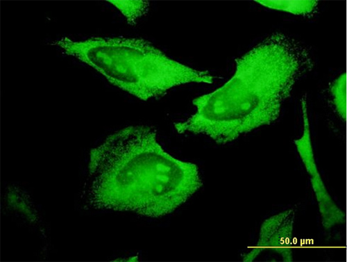
Immunofluorescence analysis of HeLa cells, staining EPCR/CD201 with ab56689 at 10 µg/ml.