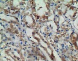
ab85086, at a 1/100 dilution, staining ENO1 in the cytoplasm and membrane of formalin fixed, paraffin embedded renal tubules by Immunohistochemistry.
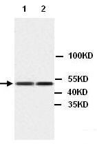
All lanes : Anti-ENO1 antibody (ab85086) at 1/200 dilutionLane 1 : MCF7 cell lysateLane 2 : Jurkat cell lysate
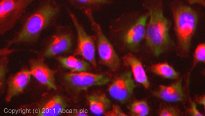
ICC/IF image of ab85086 stained HeLa cells. The cells were 4% formaldehyde fixed (10 min) and then incubated in 1%BSA / 10% normal goat serum / 0.3M glycine in 0.1% PBS-Tween for 1h to permeabilise the cells and block non-specific protein-protein interactions. The cells were then incubated with the antibody (ab85086, 1µg/ml) overnight at +4°C. The secondary antibody (green) was Alexa Fluor® 488 goat anti-rabbit IgG (H+L) used at a 1/1000 dilution for 1h. Alexa Fluor® 594 WGA was used to label plasma membranes (red) at a 1/200 dilution for 1h. DAPI was used to stain the cell nuclei (blue) at a concentration of 1.43µM.
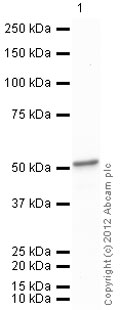
Anti-ENO1 antibody (ab85086) at 1/1000 dilution + Human ENO1 full length protein (ab89248) at 0.1 µgSecondaryGoat Anti-Rabbit IgG H&L (HRP) preadsorbed (ab97080) at 1/5000 dilutiondeveloped using the ECL techniquePerformed under reducing conditions.Exposure time : 30 seconds