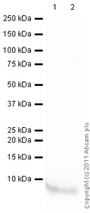
All lanes : Anti-EGF antibody (ab9695) at 0.2 µg/mlLane 1 : Human EGF full length protein (ab55483) at 0.1 µgLane 2 : Human EGF full length protein (ab55483) at 0.01 µgSecondaryGoat Anti-Rabbit IgG H&L (HRP) preadsorbed (ab97080) at 1/5000 dilutiondeveloped using the ECL techniquePerformed under reducing conditions.Exposure time : 1 minute

ab9695 at 1.0 µg/ml staining normal human skin by IHC-P. The antibody was incubated overnight at 4˚C. An HRP-labeled polymer detection system was used with a non-alcohol soluble AEC chromogen and a proteinase K antigen retrieval.
![Standard Curve for EGF (Analyte: ab51082) dilution range 1pg/ml to 1ug/ml using Capture Antibody Mouse monoclonal [S-177] to EGF (ab18635) at 5ug/ml and Detector Antibody Rabbit polyclonal to EGF (ab9695) at 0.5ug/ml](http://www.bioprodhub.com/system/product_images/ab_products/2/sub_2/12071_EGF-Primary-antibodies-ab9695-4.jpg)
Standard Curve for EGF (Analyte: ab51082) dilution range 1pg/ml to 1ug/ml using Capture Antibody Mouse monoclonal [S-177] to EGF (ab18635) at 5ug/ml and Detector Antibody Rabbit polyclonal to EGF (ab9695) at 0.5ug/ml
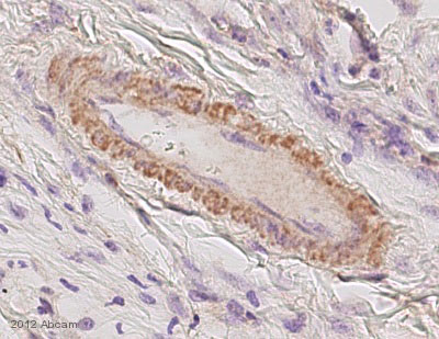
Immunohistochemical analysis of murine forepaw vessel, staining EGF with ab9695.Tissue was fixed with formaldehyde and blocked with 1% blocking solution for 15 minutes at room temperature; antigen retrieval was by proteinase K. Samples were incubated with primary antibody (4 µg/ml in BSA in TBS) for 1 hour. An undiluted HRP-conjugated goat anti-rabbit polyclonal IgG was used as the secondary antibody.See Abreview