Anti-EFHD2 antibody
| Name | Anti-EFHD2 antibody |
|---|---|
| Supplier | Abcam |
| Catalog | ab87006 |
| Prices | $370.00 |
| Sizes | 100 µl |
| Host | Rabbit |
| Clonality | Polyclonal |
| Isotype | IgG |
| Applications | WB IHC-P |
| Species Reactivities | Human, Mouse, Rat, Bovine, Pig |
| Antigen | Synthetic peptide corresponding to a region within the N terminal amino acids 2-51 (ATDELATKLS RRLQMEGEGG GETPEQPGLN GAAAAAAGAP DEAAEALGSA) of Human EFHD2, NP_077305 |
| Description | Rabbit Polyclonal |
| Gene | EFHD2 |
| Conjugate | Unconjugated |
| Supplier Page | Shop |
Product images
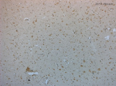 ab87006 staining EFHD2 in Human brain tissue sections by Immunohistochemistry (IHC-P - paraformaldehyde-fixed, paraffin-embedded sections). Tissue was fixed with formaldehyde and blocked with 5% serum for 30 minutes at 20°C; antigen retrieval was by heat mediation in 10mM citrate pH6, 0.1% Tween 20. Samples were incubated with primary antibody (diluted in 0.1% TritonX-100 in PBS) for 16 hours at 4°C. An undiluted HRP-conjugated Horse anti-rabbit polyclonal was used as the secondary antibody.See Abreview
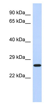 Anti-EFHD2 antibody (ab87006) at 1 µg/ml + fetal lung lysate at 10 µgSecondaryHRP conjugated anti-Rabbit IgG at 1/50000 dilution