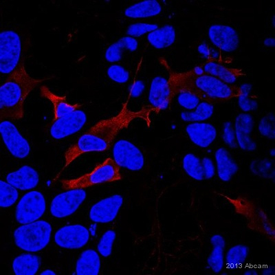
ab24368 staining EFHD2 in Human neuroblastoma cells by ICC/IF (Immunocytochemistry/immunofluorescence). Cells were fixed with formaldehyde, permeabilized with 0.1% Triton X-100 and blocked with 5% serum for 30 minutes at room temperature. Samples were incubated with primary antibody (1/100 in PBS + 5% serum) for 1 hour. DyLight®594-conjugated Mouse anti-goat IgG polyclonal (7µg/ml) was used as the secondary antibody.See Abreview
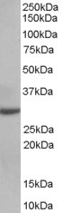
Anti-EFHD2 antibody (ab24368) at 0.03 µg/ml + Human Thymus lysate (35µg protein in RIPA buffer) at 35 µgdeveloped using the ECL techniqueObserved band size : 27 kDa (why is the actual band size different from the predicted?)

Ab24368 (0.3ug/ml) staining human EFHD2 in human tonsil by immunohistochemistry using paraffin embedded tissue. Microwaved antigen retrieval with citrate buffer pH6, HRP-staining. Positive cells A) in the interfollicular area and surrounding a vessel, B) in proximity of the epithelial criptae.