
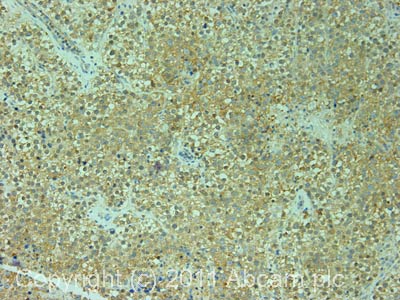
Immunohistochemistry (Formalin/PFA-fixed paraffin-embedded sections) analysis of human colon carcinoma tissue labelling EEF1G with ab72368 at 1/200 (1µg/ml). Detection: DAB.

All lanes : Anti-EEF1G antibody (ab72368) at 0.04 µg/mlLane 1 : Whole cell lysate from Hela cells at 50 µgLane 2 : Whole cell lysate from Hela cells at 15 µgLane 3 : Whole cell lysate from Hela cells at 5 µgLane 4 : Whole cell lysate from 293T cells at 50 µgLane 5 : Whole cell lysate from NIH3T3 cells at 50 µgdeveloped using the ECL technique

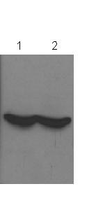
Immunoprecipitation/ Western Blot of EEF1G. Lane 1: ab72368 at 3µg/mg whole cell lysate. Lane 2: Control IgG. ab72368 at 1µg/ml for WB. Whole cell lysate from Hela cells at 1mg for IP, 20% of IP loaded. Chemiluminescence with an exposure time of 30 seconds.
All lanes : Anti-EEF1G antibody (ab72368) at 1/3000 dilution (in PBS-T for 16 hours at 4°C)Lane 1 : Monkey kidney whole cell lysate overexpressing EEF1GLane 2 : COS7 whole cell lysateLysates/proteins at 20 µg per lane.SecondaryAn HRP-conjugated Goat anti-rabbit IgG polyclonal at 1/3000 dilutiondeveloped using the ECL techniquePerformed under reducing conditions.
IHC image of ab72368 staining in human normal testis formalin fixed paraffin embedded tissue section, performed on a Leica BondTM system using the standard protocol F. The section was pre-treated using heat mediated antigen retrieval with sodium citrate buffer (pH6, epitope retrieval solution 1) for 20 mins. The section was then incubated with ab72368, 1µg/ml, for 15 mins at room temperature and detected using an HRP conjugated compact polymer system. DAB was used as the chromogen. The section was then counterstained with haematoxylin and mounted with DPX.For other IHC staining systems (automated and non-automated) customers should optimize variable parameters such as antigen retrieval conditions, primary antibody concentration and antibody incubation times.
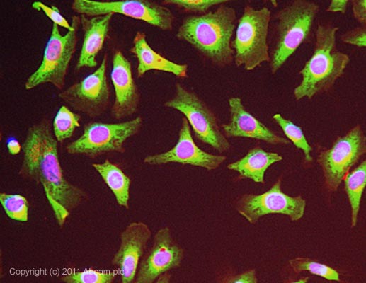
ICC/IF image of ab72368 stained HeLa cells. The cells were 4% formaldehyde fixed (10 min) and then incubated in 1%BSA / 10% normal goat serum / 0.3M glycine in 0.1% PBS-Tween for 1h to permeabilise the cells and block non-specific protein-protein interactions. The cells were then incubated with the antibody (ab72368, 1µg/ml) overnight at +4°C. The secondary antibody (green) was ab96899, DyLight® 488 goat anti-rabbit IgG (H+L) used at a 1/250 dilution for 1h.Alexa Fluor® 594 WGA was used to label plasma membranes (red) at a 1/200 dilution for 1h. DAPI was used to stain the cell nuclei (blue) at a concentration of 1.43µM.