Phospho-ATM (Ser1981) (D25E5) Rabbit mAb
| Name | Phospho-ATM (Ser1981) (D25E5) Rabbit mAb |
|---|---|
| Supplier | Cell Signaling Technology |
| Catalog | 13050 |
| Prices | $287.00 |
| Sizes | 100 µl (10 western blots) |
| Host | Rabbit |
| Clonality | Monoclonal |
| Isotype | IgG |
| Clone | D25E5 |
| Applications | WB FC |
| Species Reactivities | Human, Monkey |
| Antigen | Monoclonal antibody is produced by immunizing animals with a synthetic phosphopeptide corresponding to residues surrounding Ser1981 of human ATM protein. |
| Description | Rabbit Monoclonal |
| Gene | ATM |
| Supplier Page | Shop |
Product images
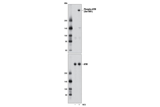 Western blot analysis of extracts from HCT 116 cells, untreated (-) or treated with neocarzinostatin (NCS 10 μM, 1 hr; +), using Phospho-ATM (Ser1981) (D25E5) Rabbit mAb (upper) and ATM (D2E2) Rabbit mAb #2873 (lower).
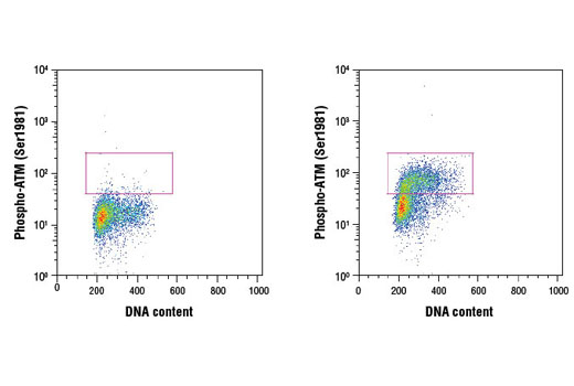 Flow cytometric analysis of HeLa cells, untreated (left) or treated with camptothecin (1 μM, 2 hr; right), using Phospho-ATM (Ser1981) (D25E5) Rabbit mAb and Propidium Iodide (PI)/RNase Staining Solution #4087 to measure DNA content. Anti-rabbit IgG (H+L), F(ab') 2 Fragment (Alexa Fluor ® 488 Conjugate) #4412 was used as a secondary antibody.